Korean J Sports Med.
2019 Dec;37(4):178-183. 10.5763/kjsm.2019.37.4.178.
Relationship between Hamstring Muscle Thickness and Knee Flexion Torque and Rate of Torque Development
- Affiliations
-
- 1Department of Kinesiology, Inha University, Busan, Korea.
- 2Department of Physical Education, Dong-A University, Busan, Korea. hjun@dau.ac.kr
- KMID: 2466808
- DOI: http://doi.org/10.5763/kjsm.2019.37.4.178
Abstract
- PURPOSE
The purpose of this study was to examine the relationships between hamstring muscle thickness and knee flexion peak torque, and rate of torque development (RTD) calculated during 0-50 ms (RTD50) and 0-200 ms (RTD200).
METHODS
Thirty-six active individuals' dominant side hamstring thickness were measured using portable ultrasound device. Participants performed maximal isometric voluntary contraction (MVIC) of knee flexion. Peak torque was identified as the maximum torque during MVIC testing. RTD was calculated initial 50 ms and 200 ms after the onset of joint torque. Pearson's correlation (r) coefficients were utilized to assess relationships between muscle thickness and knee flexion peak torque, RTD50 and RTD200. The significant level of hypothesis verification is set-up as α=0.05.
RESULTS
Greater peak torque and RTD200 was associated with greater muscle thickness of semitendinosus and semimembranosus (p<0.05). Greater RTD50 was associated with greater muscle thickness of semitendinosus only. Biceps femoris thickness was not associated with knee flexion peak torque, RTD50, and RTD200.
CONCLUSION
These results suggest that the training specific hamstring muscle (medial hamstrings) for improving muscle thickness would be effective for increasing knee flexion peak torque and RTD.
MeSH Terms
Figure
-
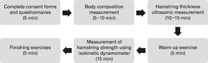
Fig. 1 Experimental design.
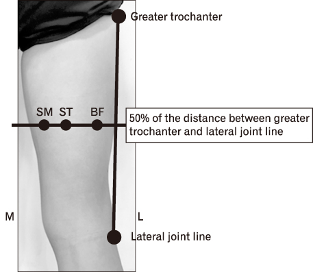
Fig. 2 Hamstring muscle thickness measurement location. SM: semimembranosus, ST: semitendinosus, BF: biceps femoris, M: medial, L: lateral.
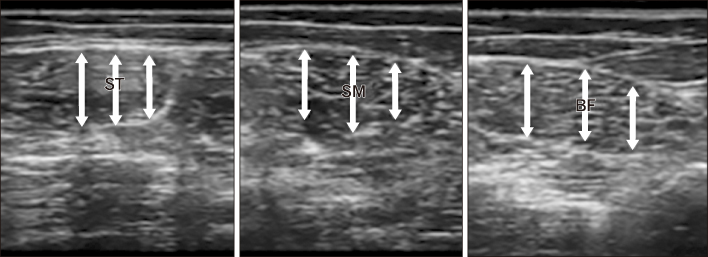
Fig. 3 Measurement of hamstring muscle thickness using Image J. ST: semitendinosus, SM: semimembranosus, BF: biceps femoris.
Reference
-
1. Lockie RG, Murphy AJ, Schultz AB, Knight TJ, Janse de. The effects of different speed training protocols on sprint acceleration kinematics and muscle strength and power in field sport athletes. J Strength Cond Res. 2012; 26:1539–1550.2. Askling C, Karlsson J, Thorstensson A. Hamstring injury occurrence in elite soccer players after preseason strength training with eccentric overload. Scand J Med Sci Sports. 2003; 13:244–250.3. Seo TB, Kim TW, Song HS, Kim YS. Comparative analysis of world class national male judo players' athletic performance related physical fitness factors. Exerc Sci. 2014; 23:171–179.4. Hunter JP, Marshall RN, McNair PJ. Relationships between ground reaction force impulse and kinematics of sprintrunning acceleration. J Appl Biomech. 2005; 21:31–43.5. Kuitunen S, Komi PV, Kyrolainen H. Knee and ankle joint stiffness in sprint running. Med Sci Sports Exerc. 2002; 34:166–173.6. Kyrolainen H, Belli A, Komi PV. Biomechanical factors affecting running economy. Med Sci Sports Exerc. 2001; 33:1330–1337.7. Krommes K, Petersen J, Nielsen MB, Aagaard P, Holmich P, Thorborg K. Sprint and jump performance in elite male soccer players following a 10-week Nordic hamstring exercise protocol: a randomised pilot study. BMC Res Notes. 2017; 10:669.8. Aagaard P, Andersen JL, Dyhre-Poulsen P, et al. A mechanism for increased contractile strength of human pennate muscle in response to strength training: changes in muscle architecture. J Physiol. 2001; 534(Pt 2):613–623.9. Aagaard P, Simonsen EB, Andersen JL, Magnusson P, Dyhre-Poulsen P. Increased rate of force development and neural drive of human skeletal muscle following resistance training. J Appl Physiol (1985). 2002; 93:1318–1326.10. Huston LJ, Wojtys EM. Neuromuscular performance characteristics in elite female athletes. Am J Sports Med. 1996; 24:427–436.11. Kakihana W, Suzuki S. The EMG activity and mechanics of the running jump as a function of takeoff angle. J Electromyogr Kinesiol. 2001; 11:365–372.12. Tillin NA, Jimenez-Reyes P, Pain MT, Folland JP. Neuromuscular performance of explosive power athletes versus untrained individuals. Med Sci Sports Exerc. 2010; 42:781–790.13. de Ruiter CJ, Van Leeuwen D, Heijblom A, Bobbert MF, de Haan A. Fast unilateral isometric knee extension torque development and bilateral jump height. Med Sci Sports Exerc. 2006; 38:1843–1852.14. van Melick N, Meddeler BM, Hoogeboom TJ, Nijhuis-van der, van Cingel RE. How to determine leg dominance: the agreement between self-reported and observed performance in healthy adults. PLoS One. 2017; 12:e0189876.15. Ruas CV, Pinto RS, Lima CD, Costa PB, Brown LE. Testretest reliability of muscle thickness, echo-intensity and cross sectional area of quadriceps and hamstrings muscle groups using B-mode ultrasound. Int J Kinesiol Sports Sci. 2017; 5:35–41.16. Mendiguchia J, Martinez-Ruiz E, Morin JB, et al. Effects of hamstring-emphasized neuromuscular training on strength and sprinting mechanics in football players. Scand J Med Sci Sports. 2015; 25:e621–e629.17. Higashihara A, Nagano Y, Ono T, Fukubayashi T. Differences in hamstring activation characteristics between the acceleration and maximum-speed phases of sprinting. J Sports Sci. 2018; 36:1313–1318.18. Opar DA, Williams MD, Timmins RG, Dear NM, Shield AJ. Knee flexor strength and bicep femoris electromyographical activity is lower in previously strained hamstrings. J Electromyogr Kinesiol. 2013; 23:696–703.
- Full Text Links
-
- Actions
-
Cited
- CITED
-
- Close
- Share
-
- Similar articles
-
- Isokinetic Strength of Quadriceps and Hamstring, Hamstrings/Quadriceps Ratios in Elite Sports Athletes
- Flexor Weakness and Clinical Outcomes after Hamstring Harvest in Anterior Cruciate Ligament Reconstruction: Comparison of Single- and Double-Tendon Harvesting Techniques
- Analysis of Torque Curve in Isokinetic Knee Dynamometer
- Comparison of Peak Torque according to Size of the Rotator Cuff Tear Patients
- Evaluation of Muscle Strength Using Isokinetic Testing and Functional Result after Total Knee Arthroplasty
