Bilateral multiple renal arteries with an extra-aortic origin and quadruple testicular veins
- Affiliations
-
- 1Medical Course, Jeju National University School of Medicine, Jeju, Korea.
- 2Department of Pathology, Jeju National University School of Medicine, Jeju, Korea.
- 3Department of Anatomy, Jeju National University School of Medicine, Jeju, Korea. spyoon@jejunu.ac.kr
- 4Institute for Medical Science, Jeju National University, Jeju, Korea.
- KMID: 2466706
- DOI: http://doi.org/10.5115/acb.19.159
Abstract
- Although variations in the urogenital vessels are relatively common, a rare case of asymmetric bilateral multiple renal arteries originating not only from the aorta but also from the testicular artery was found in a 75-year-old Korean male cadaver. Three renal arteries arose from the lateral aspect of the abdominal aorta on the right side and four from the left side. Two additional renal parenchymal branches originated from the left testicular artery, accompanied by a pair of veins out of the four testicular veins on the left side. Embryological development of the urogenital vessels is of particular importance for anatomists and clinicians.
Figure
-
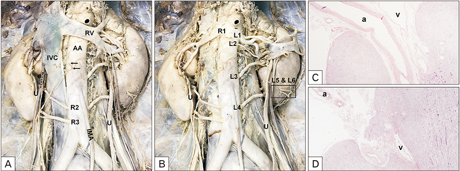
Fig. 1 Bilateral multiple renal arteries and quadruple testicular veins before (A) and after (B) reflection of the inferior vena cava (IVC). The renal arteries were designated R1 to R3 from the highest to the lowest on the right side and, similarly, L1 to L6 on the left side. R2 and R3 showed precaval courses. L5 (C) and L6 (D) originated from the left testicular artery (rectangle, B) and penetrated the renal parenchyma, which was confirmed by histopathological examination (H&E, ×100). The right testicular vein drained into the inferior vena cava (dotted arrow, A), whereas the left quadruple testicular veins drained into the left renal vein (dotted arrows, A), accompanied by L5 and L6, respectively. Arrows indicate testicular arteries. a, artery; AA, abdominal aorta; IMA, inferior mesenteric artery; RV, renal vein; U, ureter; v, vein.
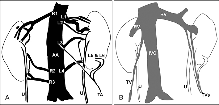
Fig. 2 Schematic drawing of the variations of the urogenital vessels. (A) Additional renal arteries arose from the abdominal aorta (AA) bilaterally and from the left testicular artery (TA). The renal arteries were designated R1 to R3 from the highest to the lowest position on the right side and, similarly, L1 to L6 on the left side. (B) The left quadruple testicular veins (TVs) drained into the left renal vein (RV), accompanied by additional left renal arteries that arose from the testicular artery. IVC, inferior vena cava; U, ureter.
Cited by 1 articles
-
Regions of the human renal artery: histomorphometric analysis
Blanca Mompeó-Corredera, Pablo Hernández-Morera, Irene Castaño-González, María del Pino Quintana-Montesdeoca, Natalia Mederos-Real
Anat Cell Biol. 2022;55(3):330-340. doi: 10.5115/acb.22.072.
Reference
-
1. Gupta R, Gupta A, Aggarwal N. Variations of gonadal veins: embryological prospective and clinical significance. J Clin Diagn Res. 2015; 9:AC08–AC10.2. Adachi B. Das Arterien system der Japaner II [The arterial system of the Japanese II]. Kyoto: Verlag der Kaiserlich-Japanischen Universität zu Kyoto;1928. p. 74–87.3. Matusz P, Miclaus G, Ples H. Study of the renal additional arteries on 1,000 CT angiography continuous series. Clin Anat. 2011; 24:408.4. Gulas E, Wysiadecki G, Szymański J, Majos A, Stefańczyk L, Topol M, Polguj M. Morphological and clinical aspects of the occurrence of accessory (multiple) renal arteries. Arch Med Sci. 2018; 14:442–453.5. Bordei P, Sapte E, Iliescu D. Double renal arteries originating from the aorta. Surg Radiol Anat. 2004; 26:474–479.6. Ozkan U, Oğuzkurt L, Tercan F, Kizilkiliç O, Koç Z, Koca N. Renal artery origins and variations: angiographic evaluation of 855 consecutive patients. Diagn Interv Radiol. 2006; 12:183–186.7. Shoja MM, Tubbs RS, Shakeri AB, Oakes WJ. Origins of the gonadal artery: embryologic implications. Clin Anat. 2007; 20:428–432.8. Ambos MA, Bosniak MA, Lefleur RS. Blood flow to the kidney via the gonadal-renal capsular artery. Urol Radiol. 1979; 1:11–16.9. Hinata N, Suzuki R, Ishizawa A, Miyake H, Rodriguez-Vazquez JF, Murakami G, Fujisawa M. Fetal development of the mesonephric artery in humans with reference to replacement by the adrenal and renal arteries. Ann Anat. 2015; 202:8–17.10. Felix W. The development of the urogenital organs. In : Keibel F, Mall FP, editors. Manual of Human Embryology. II. Philadelphia, PA: J.B. Lippincott Company;1912. p. 752–880.11. Favorito LA, Costa WS, Sampaio FJ. Applied anatomic study of testicular veins in adult cadavers and in human fetuses. Int Braz J Urol. 2007; 33:176–180.12. Nallikuzhy TJ, Rajasekhar SS, Malik S, Tamgire DW, Johnson P, Aravindhan K. Variations of the testicular artery and vein: a meta-analysis with proposed classification. Clin Anat. 2018; 31:854–869.13. Mazengenya P. Multiple variations of the renal and testicular vessels: possible embryological basis and clinical importance. Surg Radiol Anat. 2016; 38:729–733.14. Rudloff U, Holmes RJ, Prem JT, Faust GR, Moldwin R, Siegel D. Mesoaortic compression of the left renal vein (nutcracker syndrome): case reports and review of the literature. Ann Vasc Surg. 2006; 20:120–129.15. Sharma P, Salwan SK. Anomalous right testicular artery and vein: embryologic explanation and clinical implications. J Clin Diagn Res. 2011; 5:1631–1633.
- Full Text Links
-
- Actions
-
Cited
- CITED
-
- Close
- Share
-
- Similar articles
-
- Unique variation of the left testicular artery passing through a vascular hiatus in renal vein
- Abnormal ramification pattern of the renal and testicular vessels
- A rare combined variation of the coeliac trunk, renal and testicular vasculature
- Multiple Vascular Variations in Posterior Abdominal Region: A Case Report
- Case Report on Horseshoe Kidney
