J Korean Ophthalmol Soc.
2019 Dec;60(12):1339-1343. 10.3341/jkos.2019.60.12.1339.
Hemophilia A in a Female Patient with Recurrent Vitreous Hemorrhage
- Affiliations
-
- 1Department of Ophthalmology, Yonsei University Wonju College of Medicine, Wonju, Korea. Dingo84@yonsei.ac.kr
- KMID: 2466193
- DOI: http://doi.org/10.3341/jkos.2019.60.12.1339
Abstract
- PURPOSE
To report a case of recurrent intraocular hemorrhage due to type A hemophilia in a female patient without any previous medical history.
CASE SUMMARY
A 51-year-old female patient without any previous medical history was referred to our clinic due to blurred vision in her left eye. Slit lamp microscopy of the anterior segment was nonspecific. Fundus examination revealed vitreous hemorrhage with retinal tear in her left eye. Vitrectomy and cataract surgery were performed. One day after surgery, hyphema and vitreous hemorrhage recurred. A coagulation disorder was suspected and further serological evaluation was conducted. Coagulation factor analyses showed that the activity of coagulation factors 8 and 12 decreased to 25% and 47%, respectively. Genetic sequence analyses were conducted, and a missense mutation of C6724G> A] was found in exon 25, and type A hemophilia was confirmed.
CONCLUSIONS
In patients who tend to show persistent bleeding even after proper treatment, hematological evaluation including coagulation factor assays, and the possibility of rare diseases such as hemophilia should be considered.
MeSH Terms
Figure
-
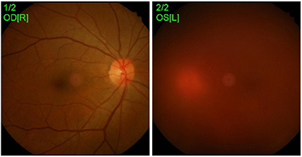
Figure 1 Fundus photo examination. At initial visit. OD = oculus dexter; R= right; OS = oculus sinister; L = left.
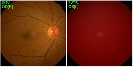
Figure 2 Fundus photo examination. After 1st vitrectomy (1 day). OD = oculus dexter; R= right; OS = oculus sinister; L = left.

Figure 3 Fundus photo examination OCT. (A) After vitrectomy (2 months). (B) After 2nd vitrectomy (1 year). OD = oculus dexter; R= right; OS = oculus sinister; L = left; OCT = optical coherence tomography.

Figure 4 Genetic sequencing of patient. Gene of F8. CDS = coding sequence; UTR = untranslated regiion; HGMD = Human Gene Mutation Database; CHAMP = The Chip Analysis Methylation Pipeline.
Reference
-
1. Yoon JT, Kim CK, Sohn JH, Yoon YH. Systemic risk factors for postoperative vitreous hemorrhage following diabetic vitrectomy. J Korean Ophthalmol Soc. 2001; 42:434–440.2. Chung A, Chin EK, Almeida DR. Recurrent vitreous hemorrhage despite pars plana vitrectomy, laser, and injections. JAMA Ophthalmol. 2016; 134:231–232.3. Hoyer LW. Hemophilia A. N Engl J Med. 1994; 330:38–47.4. Gilbert L, Paroskie A, Gailani D, et al. Haemophilia A carriers experience reduced health-related quality of life. Haemophilia. 2015; 21:761–765.5. Baglin T, Gray E, Greaves M, et al. Clinical guidelines for testing for heritable thrombophilia. Br J Haematol. 2010; 149:209–220.6. DI Michele DM, Gibb C, Lefkowitz JM, et al. Severe and moderate haemophilia A and B in US females. Haemophilia. 2014; 20:e136–e143.7. Karmaker M, Zerin I, Afrose S, et al. Acquired hemophilia in female - a case report. J Med. 2017; 18:119–122.8. Mazurkiewicz-Pisarek A, Płucienniczak G, Ciach T, Płucienniczak A. The factor VIII protein and its function. Acta Biochim Pol. 2016; 63:11–16.9. Hwang SH, Kim MJ, Lim JA, et al. Profiling of factor VIII mutations in Korean haemophilia A. Haemophilia. 2009; 15:1311–1317.10. Wagenman BL, Townsend KT, Mathew P, Crookston KP. The laboratory approach to inherited and acquired coagulation factor deficiencies. Clin Lab Med. 2009; 29:229–252.11. Gitschier J, Wood WI, Goralka TM, et al. Characterization of the human factor VIII gene. Nature. 1984; 312:326–330.12. Levinson B, Kenwrick S, Lakich D, et al. A transcribed gene in an intron of the human factor VIII gene. Genomics. 1990; 7:1–11.13. Liu ML, Shen BW, Nakaya S, et al. Hemophilic factor VIII C1- and C2-domain missense mutations and their modeling to the 1.5-angstrom human C2-domain crystal structure. Blood. 2000; 96:979–987.14. Vehar GA, Keyt B, Eaton D, et al. Structure of human factor VIII. Nature. 1984; 312:337–342.15. Aslam S, Poon MC, Yee VC, et al. Factor XIIIA calgary: a candidate missense mutation (Leu667Pro) in the beta barrel 2 domain of the factor XIIIA subunit. Br J Haematol. 1995; 91:452–457.16. Bhoi D, Kashyap L. Perioperative management of a patient with hemophilia A and crigler-najjar syndrome. J Anaesthesiol Clin Pharmacol. 2013; 29:582–584.17. Shahriari M, Bazrafshan A, Moghadam M, Karimi M. Severe hemophilia in a girl infant with mosaic Turner syndrome and persistent hyperplastic primary vitreous. Blood Coagul Fibrinolysis. 2016; 27:352–353.18. Kobayashi H, Honda Y. Intraocular hemorrhage in a patient with hemophilia. Metab Ophthalmol. 1984-1985; 8:27–30.
- Full Text Links
-
- Actions
-
Cited
- CITED
-
- Close
- Share
-
- Similar articles
-
- Successful Non-operative Management of Intra-abdominal Hemorrhage in Two Patients with Hemophilia A
- Iliacus Hematoma with Femoral Neuropathy in Hemophilia: A Case report
- An Experience of Treatment in a Case of Exophthalmos with the Hemophilia
- Experimental Vitreous Hemorrhage
- Efficacy of Intravitreal Triamcinolone Acetonide for Eyes with Postvitrectomy Diabetic Vitreous Hemorrhage
