Squamous Cell Carcinoma of the Extrahepatic Common Hepatic Duct
- Affiliations
-
- 1Department of Pathology, Gil Medical Center, Gachon University College of Medicine, Incheon, Korea. clara_nrk@gilhospital.com
- 2Department of Surgery, Gil Medical Center, Gachon University College of Medicine, Incheon, Korea.
- KMID: 2465450
- DOI: http://doi.org/10.4132/jptm.2018.09.03
Abstract
- We report a rare case of hilar squamous cell carcinoma. A 62-year-old Korean woman complaining of nausea was referred to our hospital. Her biliary computed tomography revealed a 28 mm-sized protruding solid mass in the proximal common bile duct. The patient underwent left hemihepatectomy with S1 segmentectomy and segmental excision of the common bile duct. Microscopically, the tumor was a moderately differentiated squamous cell carcinoma of the extrahepatic bile duct, without any component of adenocarcinoma or metaplastic portion in the biliary epithelium. Immunohistochemically, the tumor was positive for cytokeratin (CK) 5/6, CK19, p40, and p63. Squamous cell carcinoma of the extrahepatic bile duct is rare. To date, only 24 cases of biliary squamous cell carcinomas have been reported. Here, we provide a clinicopathologic review of previously reported extrahepatic bile duct squamous cell carcinomas.
MeSH Terms
Figure
-
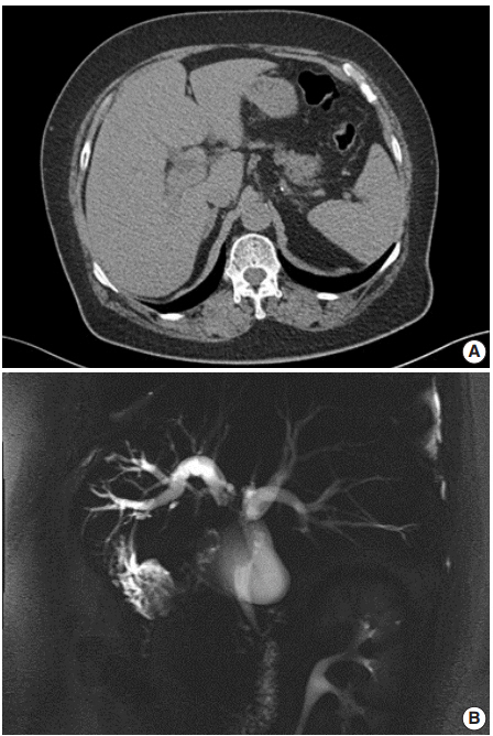
Fig. 1. (A) Computed tomography reveals perihilar cholangiocarcinoma with metastatic lymph nodes. (B) Magnetic resonance cholangiopancreatography shows strictures of the left intrahepatic duct to common hepatic duct.

Fig. 2. (A) The gross specimen revealed a protruded mass (arrows) accounting for all layers of the hepatic duct wall. (B) Histologically, thickened papillary squamous epithelium shows moderately differentiated dyskeratotic squamous cells with keratin pearls with stromal invasion. (C) Surface epithelium shows a transition from unilayered cuboidal to squamous epithelium (arrow). (D) Immunohistochemically, the tumor cells are positive for p63 (left) and p40 (right).

Fig. 3. Ultrastructurally, ovoid-shaped tumor cells have cytoplasmic tonofilaments (white arrows) and are connected with wellformed desmosomes (black arrows, ×2,500).
Reference
-
1. Cabot RC, Painter FM. Case records of the Massachusetts General Hospital: Case 16261: four months’ jaundice and rectal pain. N Eng J Med. 1930; 202:1260–2.2. Burger RE, Meeker WR, Luckett PM. Squamous cell carcinoma of the common bile duct. South Med J. 1978; 71:216–9.
Article3. Gulsrud PO, Feinberg M, Koretz RL. Rapid development of cirrhosis secondary to squamous cell carcinoma of the common bile duct. Dig Dis Sci. 1979; 24:166–9.
Article4. Aranha GV, Reyes CV, Greenlee HB, Field T, Brosnan J. Squamous cell carcinoma of the proximal bile duct: a case report. J Surg Oncol. 1980; 15:29–35.5. Kim KS, Park HB, Yeo HS, et al. A case of squamous cell carcinoma of the common bile duct. Korean J Gastrointest Endosc. 1999; 19:486–90.6. Cho T, Nakamura J, Tomita H, et al. A case of squamous cell carcinoma of the distal extrahepatic bile duct. J Jpn Sug Assoc. 2000; 61:1853–6.7. Gatof D, Chen YK, Shah RJ. Primary squamous cell carcinoma of the bile duct diagnosed by transpapillary cholangioscopy: case report and review. Gastrointest Endosc. 2004; 60:300–4.
Article8. La Greca G, Conti P, Urrico GS, et al. Biliary squamous cell carcinoma. Chir Ital. 2004; 56:289–95.9. Sewkani A, Kapoor S, Sharma S, et al. Squamous cell carcinoma of the distal common bile duct. JOP. 2005; 6:162–5.10. Abbas R, Willis J, Kinsella T, Siegel C, Sanabria J. Primary squamous cell carcinoma of the main hepatic bile duct. Can J Surg. 2008; 51:E85–6.11. Price L, Kozarek R, Agoff N. Squamous cell carcinoma arising within a choledochal cyst. Dig Dis Sci. 2008; 53:2822–5.
Article12. Kim GM, Choi GH, Kim DH, Kang CM, Lee WJ. A case of squamous cell carcinoma of the distal common bile duct. Korean J Hepatobiliary Pancreat Surg. 2008; 12:210–3.13. Yamana I, Kawamoto S, Nagao S, Yoshida T, Yamashita Y. Squamous cell carcinoma of the hilar bile duct. Case Rep Gastroenterol. 2011; 5:463–70.
Article14. Yoo Y, Mun S. Synchronous double primary squamous cell carcinoma and adenocarcinoma of the extrahepatic bile duct: a case report. J Med Case Rep. 2015; 9:116.
Article15. Goto T, Sasajima J, Koizumi K, et al. Primary poorly differentiated squamous cell carcinoma of the extrahepatic bile duct. Intern Med. 2016; 55:1581–4.
Article16. Nishiguchi R, Kim DH, Honda M, Sakamoto T. Squamous cell carcinoma of the extrahepatic bile duct with metachronous para-aortic lymph node metastasis successfully treated with S-1 plus cisplatin. BMJ Case Rep. 2016; 2016:bcr2016218177.
Article17. Yang G, Li J, Meng D. Primary squamous cell cholangiocarcinoma: a case report. Int J Clin Exp Pathol. 2016; 9:5772–6.18. Mori H, Kaneoka Y, Maeda A, Takayama Y, Fukami Y, Onoe S. A perihilar bile duct squamous cell carcinoma treated by left hepatic lobe and caudate lobe resection, subtotal stomach preserving pancreatoduodenectomy, and portal vein resection. Jpn J Gastroenterol Surg. 2017; 50:26–32.
Article19. Pastuszak M, Groszewski K, Pastuszak M, Dyrla P, Wojtun´ S, Gil J. Cytokeratins in gastroenterology: systematic review. Prz Gastroenterol. 2015; 10:61–70.
Article20. Khan SA, Davidson BR, Goldin RD, et al. Guidelines for the diagnosis and treatment of cholangiocarcinoma: an update. Gut. 2012; 61:1657–69.
Article
- Full Text Links
-
- Actions
-
Cited
- CITED
-
- Close
- Share
-
- Similar articles
-
- ERCP findings of extrahepatic bile duct carcinoma
- Obstructive Jaundice Caused by the Fragment of Hepatocellular Carcinoma in the Common Hepatic Duct Confirmed by Peroral Choledochoscopy
- A Case of Multiple Papillary Adenocarcinoma of the Extrahepatic Bile Duct : Findings of ERCP
- A Case of Squamous Cell Carcinoma of the Distal Common Bile Duct
- A Case of Squamous Cell Carcinoma of the Common Bile Duct
