Cytomorphological Features of Hyperchromatic Crowded Groups in Liquid-Based Cervicovaginal Cytology: A Single Institutional Experience
- Affiliations
-
- 1Department of Pathology, Seoul National University Hospital, Seoul National University College of Medicine, Seoul, Korea. haeryoung.kim@snu.ac.kr
- KMID: 2465439
- DOI: http://doi.org/10.4132/jptm.2019.08.14
Abstract
- BACKGROUND
Hyperchromatic crowed groups (HCGs) are defined as three-dimensional aggregates of crowded cells with hyperchromatic nuclei, and are frequently encountered in cervicovaginal liquid-based cytology (LBC). Here, we aimed to examine the prevalence of HCGs in cervicovaginal LBC and the cytomorphological characteristics of various epithelial cell clusters presenting as HCGs.
METHODS
We first examined the prevalence of HCGs in a "routine cohort" of LBC cytology (n=331), consisting of all cervicovaginal LBCs accessioned over 3 days from outpatient clinics (n=179) and the screening population (n=152). Then we examined a second "high-grade epithelial cell abnormalities (H-ECA) cohort" (n=69) of LBCs diagnosed as high-grade squamous intraepithelial lesion (HSIL), squamous cell carcinoma (SCC), or adenocarcinoma during 1 year.
RESULTS
HCGs was observed in 34.4% of the routine cohort and were significantly more frequent in the epithelial cell abnormality category compared to the non-neoplastic category (p=.003). The majority of HCGs represented atrophy (70%). Of the 69 histologically confirmed H-ECA cases, all contained HCGs. The majority of cases were HSIL (62%), followed by SCC (16%). Individually scattered neoplastic cells outside the HCGs were significantly more frequent in SCCs compared to glandular neoplasia (p=.002). Despite the obscuring thick nature of the HCGs, examining the edges and the different focal planes of the HCGs and the background were helpful in defining the nature of the HCGs.
CONCLUSIONS
HCGs were frequently observed in cervicovaginal LBC and were mostly non-neoplastic; however, neoplastic HCGs were mostly high-grade lesions. Being aware of the cytomorphological features of different HCGs is important in order to avoid potential false-negative cytology interpretation.
MeSH Terms
Figure
-
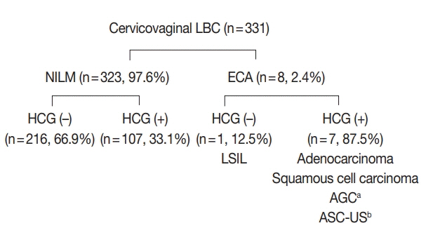
Fig. 1. Summary of the routine cervicovaginal liquid-based cytology (LBC) cases. Hyperchromatic crowed groups (HCGs) were significantly more frequent in the epithelial cell abnormality (ECA) category compared to the negative for intraepithelial lesion or malignancy (NILM) category, and neoplastic HCGs were high-grade lesions. LSIL, low-grade squamous intraepithelial lesion; AGC, atypical glandular cell; ASC-US, atypical squamous cells of uncertain significance. a Subsequently diagnosed as adenocarcinoma; b Only nonneoplastic HCGs were observed.
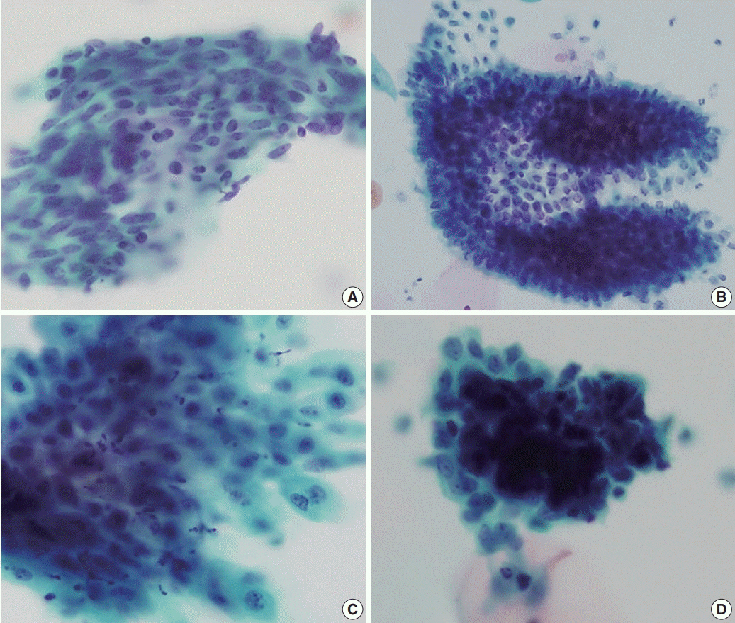
Fig. 2. Cytological features of non-neoplastic hyperchromatic crowed group (HCG). (A) Atrophic HCGs consisted of bland-looking parabasal cells with streaming patterns. (B) Regular honeycomb arrangement of endocervical cells. (C) Reactive endocervical cells demonstrating mild nuclear size variation and nucleoli but smooth nuclear membranes. (D) Biphasic pattern observed in endometrial cell clusters.

Fig. 3. Summary of the high-grade epithelial cell abnormalities cohort. The majority were squamous lesions (78%).

Fig. 4. Cytological features of neoplastic hyperchromatic crowed group (HCG). (A–C) HCGs of squamous cell carcinoma demonstrate dense cellular clusters with chaotic arrangement, and the individual cells are markedly hyperchromatic. The periphery of the HCG is flattened. When present, tumor diathesis and scattered dyskeratotic cells are helpful features in recognizing squamous cell carcinomas (C). (D, E) HCGs of endocervical adenocarcinomas. A vague acinar arrangement is seen within the HCG with feathering of the border (D). Smaller strips and rosettes of tumor cells are helpful clues in the diagnosis (E). (F) HCGs of well-differentiated endometrioid carcinoma were tight clusters of neoplastic cells that were smaller than endocervical adenocarcinoma. More obvious nuclear atypia were present in high-grade tumors and intracytoplasmic neutrophils were also present (inset).
Cited by 1 articles
-
Cytopathologic features of human papillomavirus–independent, gastric-type endocervical adenocarcinoma
Min-Kyung Yeo, Go Eun Bae, Dong-Hyun Kim, In-Ock Seong, Kwang-Sun Suh
J Pathol Transl Med. 2022;56(5):260-269. doi: 10.4132/jptm.2022.07.05.
Reference
-
1. DeMay RM. Common problems in Papanicolaou smear interpretation. Arch Pathol Lab Med. 1997; 121:229–38.2. Chivukula M, Austin RM, Shidham VB. Evaluation and significance of hyperchromatic crowded groups (HCG) in liquid-based paps. Cytojournal. 2007; 4:2.3. Croll E, Rana DN, Walton LJ. Hyperchromatic crowded cell groups in gynaecological liquid-based cytology samples. Br J Biomed Sci. 2010; 67:154–63.
Article4. Gupta N, John D, Dudding N, Crossley J, Smith JH. Factors contributing to false-negative and potential false-negative cytology reports in SurePath liquid-based cervical cytology. Cytopathology. 2013; 24:39–43.
Article5. Oh EJ, Jung CK, Kim DH, et al. Current cytology practices in Korea: a nationwide survey by the Korean Society for Cytopathology. J Pathol Transl Med. 2017; 51:579–87.
Article6. Lim SC, Yoo CW. Current status of and perspectives on cervical cancer screening in Korea. J Pathol Transl Med. 2019; 53:210–6.
Article7. Feratovic R, Lewin SN, Sonoda Y, et al. Cytologic findings after fertility-sparing radical trachelectomy. Cancer. 2008; 114:1–6.
Article8. Diaz-Rosario LA, Kabawat SE. Cell block preparation by inverted filter sedimentation is useful in the differential diagnosis of atypical glandular cells of undetermined significance in ThinPrep specimens. Cancer. 2000; 90:265–72.
Article9. Ge Y, Mody DR, Smith D, Anton R. p16(INK4a) and ProEx C immunostains facilitate differential diagnosis of hyperchromatic crowded groups in liquid-based Papanicolaou tests with menstrual contamination. Acta Cytol. 2012; 56:55–61.10. Evered A, Edwards J, Powell G. Image analysis of hyperchromatic crowded cell groups in SurePath cervical cytology. Cytopathology. 2013; 24:113–22.
Article
- Full Text Links
-
- Actions
-
Cited
- CITED
-
- Close
- Share
-
- Similar articles
-
- The Usefulness of Cervicovaginal Cytology as a Primary Screening Test
- Liquid-Based Cytology in Fine-Needle Aspirates of the Thyroid and Breast
- A Case of Metastatic Angiosarcoma Diagnosed by Liquid-Based Preparation: Peculiar Cytoplasmic Changes
- Evaluation of the Manual Method of Liquid-Based Uterine Cervicovaginal Cytology - By The Manual Method Based on SurePathTM Methodology
- A Comparision of Surepath(TM) Liquid-Based Smear with a Conventional Smear for Cervicovaginal Cytology-with Reference to a Histological Diagnosis
