The Role of digital subtraction angiography in the ventricular spot sign on the computed tomography angiography
- Affiliations
-
- 1Department of Neurosurgery, Deajeon St. Mary's Hospital, College of Medicine, The Catholic University of Korea, Daejeon, Republic of Korea. tkddnr79@hanmail.net
- 2Department of Neurosurgery, St. Vincent's Hospital, College of Medicine, The Catholic University of Korea, Suwon, Republic of Korea.
- KMID: 2465012
- DOI: http://doi.org/10.7461/jcen.2019.21.1.24
Abstract
OBJECTIVE
The spot sign on computed tomography angiography is little known about the relationship between the spot sign and the results of cerebral angiography We retrospectively analyzed the spot sign, digital subtraction angiography results, and other factors. MATERIAL AND METHODS: From December 2009 to May 2014, DSA was performed in 52 ICH patients with non-specific location or abnormalities on CTA findings. 26 of those patients, whose initial CTA showed the spot sign, were analyzed. Two groups, one with the spot sign in the ventricle (Group A) and others with the spot sign in another location (Group B) were statistically compared.
RESULTS
The mean age of the study subjects was 46.9 years (range, 15 to 80 years) and the percentage of males was 53.8%. Thirteen of 26 patients had ICH without intraventricular hemorrhage, and 6 patients had co-existing IVH. In 17 cases, the DSA results were negative. Seven patients were diagnosed with pseudoaneurysms, and two cases showed developmental venous anomalies. Group A consisted of the 8 patients (30.8%) who showed the spot sign in a ventricle. The number of pseudoaneurysms was statistically significantly higher in Group A than in Group B (71.4% versus 28.6%; OR, 13.3; 95% CI, 1.7-103.8 P = 0.014). All three patients who underwent endovascular treatment were members of Group A (P = 0.022), whereas most (92.3%) of those in Group B underwent surgical evacuation. (P = 0.030).
CONCLUSION
When CTA shows the spot sign in a ventricle, it is a clue that an existing underlying vascular lesion requires endovascular treatment.
MeSH Terms
Figure
-

Fig. 1 A 54-year-old male (A) Non-enhanced computed tomography (NECT) shows a left frontal ICH. (B) Hyperdensity apparent on a post contrast CT image (black arrow), called the ‘spot sign,’ is detected. There is no definite vascular abnormality on DSA. (C) A follow-up brain CT shows expansion of the hematoma

Fig. 2 A 46-year-old female with a developmental venous anomaly. (A) A NECT demonstrate ICH in the left cerebellum. (B) The CTA shows the ‘spot sign’ (black arrow) within the hematoma. (C) An umbrella-shaped collection of dilated medullary veins, the so-called ‘caput medusae’ with a dilated transcortical vein (white arrow) is detected during the venous phase of the DSA.
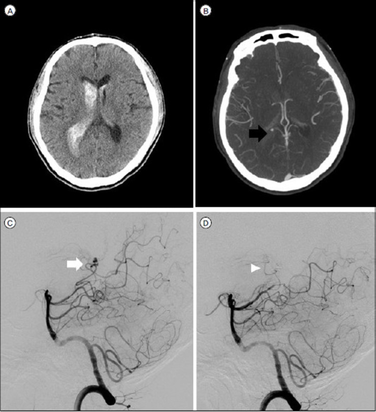
Fig. 3 A 58-year-old male with a pseudoaneurysm (A) NECT demonstrate pure IVH (B) CTA shows the spot sign (black arrow) in the right lateral ventricle. (C) A vertebral angiogram (lateral view) shows a pseudoaneurysm (white arrow) in the right posterior choroidal artery. (D) After embolization with glue, there is no more contrast filling in the pseudoaneurysm on DSA (White arrowhead).
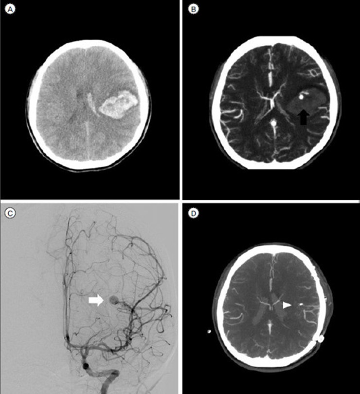
Fig. 4 A 15-year-old female with a pseudoaneurysm (A) NECT shows ICH in the right frontal lobe with IVH. (B) CTA shows the spot sign (black arrow) within the hematoma. (C) DSA (AP view) reveals an AVM with a pseudoaneurysm (white arrow). (D) After surgical treatment, there is no hematoma or pseudoaneurysm that showed contrast filling on the CTA MIP image (the white arrowhead is a post-operation hemo-clip).
Reference
-
1. Brzozowski K, Frankowska E, Piasecki P, Ziecina P, Zukowski P, Boguslawska-Walecka R. The use of routine imaging data in diagnosis of cerebral pseudoaneurysm prior to angiography. Eur J Radiol. 2011; 12. 80(3):e401–e409. PMID: 21227615.
Article2. Ciceri EF, Regna-Gladin C, Erbetta A, Chiapparini L, Nappini S, Savoiardo M, et al. Iatrogenic intracranial pseudoaneurysms: neuroradiological and therapeutical considerations, including endovascular options. Neurol Sci. 2006; 11. 27(5):317–322. PMID: 17122940.
Article3. Davis SM, Broderick J, Hennerici M, Brun NC, Diringer MN, Mayer SA, et al. Hematoma growth is a determinant of mortality and poor outcome after intracerebral hemorrhage. Neurology. 2006; 4. 66(8):1175–1181. PMID: 16636233.
Article4. de Andrade AF, Figueiredo EG, Caldas JG, Paiva WS, De Amorim RL, Puglia P, et al. Intracranial vascular lesions associated with small epidural hematomas. Neurosurgery. 2008; 2. 62(2):416–420. discussion 20–1. PMID: 18382319.
Article5. Demchuk AM, Dowlatshahi D, Rodriguez-Luna D, Molina CA, Blas YS, Dzialowski I, et al. Prediction of haematoma growth and outcome in patients with intracerebral haemorrhage using the CT-angiography spot sign (PREDICT): a prospective observational study. The Lancet Neurology. 2012; 11(4):307–314. PMID: 22405630.
Article6. Dowlatshahi D, Wasserman JK, Momoli F, Petrcich W, Stotts G, Hogan M, et al. Evolution of computed tomography angiography spot sign is consistent with a site of active hemorrhage in acute intracerebral hemorrhage. Stroke. 2014; 1. 45(1):277–280. PMID: 24178918.
Article7. Gazzola S, Aviv RI, Gladstone DJ, Mallia G, Li V, Fox AJ, et al. Vascular and nonvascular mimics of the CT angiography “spot sign” in patients with secondary intracerebral hemorrhage. Stroke. 2008; 4. 39(4):1177–1183. PMID: 18292380.
Article8. Heck O, Anxionnat R, Lacour JC, Derelle AL, Ducrocq X, Richard S, et al. Rupture of lenticulostriate artery aneurysms. J Neurosurg. 2014; 2. 120(2):426–433. PMID: 24053505.
Article9. Hemphill JC 3rd, Greenberg SM, Anderson CS, Becker K, Bendok BR, Cushman M, et al. Guidelines for the Management of Spontaneous Intracerebral Hemorrhage: A Guideline for Healthcare Professionals From the American Heart Association/American Stroke Association. Stroke. 2015; 7. 46(7):2032–2060. PMID: 26022637.
Article10. Ito H, Morishima H, Onodera H, Wakui D, Uchida M, Sase T, et al. Acute phase endovascular intervention on a pseudoaneurysm formed due to rupture of an anterior communicating artery aneurysm. J Neurointerv Surg. 2015; 3. 7(3):e9. PMID: 24565758.
Article11. Lei C, Wu B, Liu M, Cao T, Wang Q, Dong W. Differences Between Vascular Structural Abnormality and Hypertensive Intracerebral Hemorrhage. J Stroke Cerebrovasc Dis. 2015; 8. 24(8):1811–1816. PMID: 26077131.
Article12. McLaughlin MR, Kondziolka D, Flickinger JC, Lunsford S, Lunsford LD. The prospective natural history of cerebral venous malformations. Neurosurgery. 1998; 8. 43(2):195–200. discussion 200–1. PMID: 9696070.
Article13. Murias Quintana E, Gil Garcia A, Vega Valdes P, Meilan Martinez A, Botana Fernandez M, Gutierrez Morales JC, et al. [Our experience in the diagnosis and treatment of cerebral pseudoaneurysms]. Radiologia. 2012; Jan-Feb. 54(1):65–72. PMID: 21641006.
Article14. Naff NJ, Wemmer J, Hoenig-Rigamonti K, Rigamonti DR. A longitudinal study of patients with venous malformations: documentation of a negligible hemorrhage risk and benign natural history. Neurology. 1998; 6. 50(6):1709–1714. PMID: 9633715.
Article15. Rammos SK, Maina R, Lanzino G. Developmental venous anomalies: current concepts and implications for management. Neurosurgery. 2009; 7. 65(1):20–29. discussion 9–30. PMID: 19574822.16. Romero JM, Kelly HR, Delgado Almandoz JE, Hernandez-Siman J, Passanese JC, Lev MH, et al. Contrast extravasation on CT angiography predicts hematoma expansion and mortality in acute traumatic subdural hemorrhage. AJNR Am J Neuroradiol. 2013; 8. 34(8):1528–1534. PMID: 23449655.
Article17. Tan LA, Kasliwal MK, Johnson AK, Lopes DK. The “Spot Sign” secondary to a ruptured lenticulostriate artery aneurysm. Clin Imaging. 2014; Jul-Aug. 38(4):508–509. PMID: 24629789.
Article
- Full Text Links
-
- Actions
-
Cited
- CITED
-
- Close
- Share
-
- Similar articles
-
- Radiologic consideration of intra-arterial digital subtraction angiography
- Multimodal Imaging Follow-up of a Thrombosed Developmental Venous Anomaly: CT, CT Angiography and Digital Subtraction Angiography
- A Clinical Significance of lntraarterial Digital Subtraction Angiography in Renal Diseases
- Spot Sign on Initial Brain Computed Tomography Angiography Source Image to Predict Large Hemorrhagic Transformation after Middle Cerebral Artery Infarction
- Rupture of De Novo Anterior Communicating Artery Aneurysm 8 Days after the Clipping of Ruptured Middle Cerebral Artery Aneurysm
