Biomechanical Properties of the Cranial Dura Mater with Puncture Defects: An In Vitro Study
- Affiliations
-
- 1Department of Neurosurgery, Kutahya Health Science University, Kutahya, Turkey.
- 2Department of Neurosurgery, Dokuz Eylul University School of Medicine, Izmir, Turkey. ceren.kizmazoglu@gmail.com
- 3Department of Biomechanics, Dokuz Eylul University School of Medicine Health Science Institute, Izmir, Turkey.
- 4Department of Pathology, Forensic Medicine Institution , Izmir, Turkey.
- 5Department of Neurosurgery, EskiÅŸehir Osmangazi University School of Medicine, Eskisehir, Turkey.
- KMID: 2463668
- DOI: http://doi.org/10.3340/jkns.2018.0130
Abstract
OBJECTIVE
The primary aim of this investigation was to explore the nature of dura mater biomechanics following the introduction of puncture defect(s).
METHODS
Twenty-eight dura mater specimens were collected during autopsy from the department of forensic medicine of the authors' institution. Specimens were divided randomly into one of four groups : group I (cranial dura mater; n=7), group II (cranial dura mater with one puncture defect; n=7); group III (cranial dura mater with two puncture defects; n=7), and group IV (cranial dura mater with three puncture defects; n=7).
RESULTS
The mean±standard deviation tensile strengths of the dura mater were 8.35±3.16, 8.22±3.32, 7.13±1.77, and 6.94±1.93 MPa for groups I, II, III, and IV, respectively. There was no statistical difference between all groups. A single, two or more punctures of the dura mater using a 20-gauge Quincke needle did not affect cranial dura tensile strength.
CONCLUSION
This biomechanical study may contribute to the future development of artificial dura mater substitutes and medical needles that have a lower negative impact on the biomechanical properties of dura mater.
MeSH Terms
Figure
-
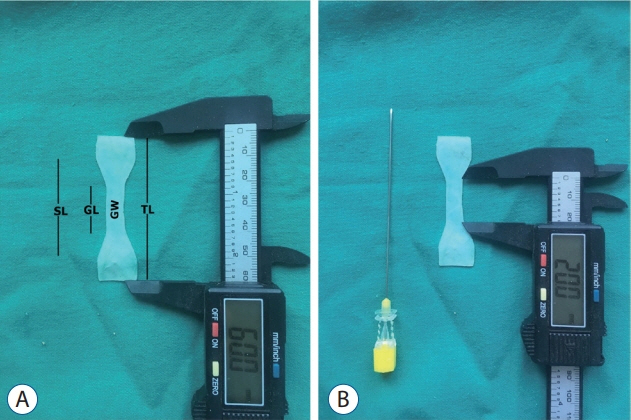
Fig. 1. A : Dimensions of the cranial dura mater specimens showing. B : Dimensions of the 20 G Quincke needle (Egemen International Inc., Izmir, Turkey). SL : shoulder length, GL : gauge length, GW : gauge width, TL : total length.

Fig. 2. A and B : The bevel tip needle stylet and needle cannula.
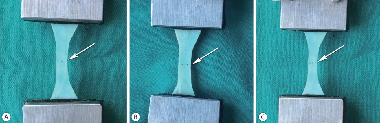
Fig. 3. Puncture defects before the uniaxial tension test. A : One-puncture defect (arrow). B : Two-puncture defects (arrow). C : Three puncture defects (arrow).
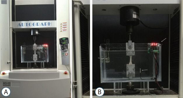
Fig. 4. The test setup (A) and white arrows show (B, left to right) heating element, waterproof sensor, digital thermometer controller.
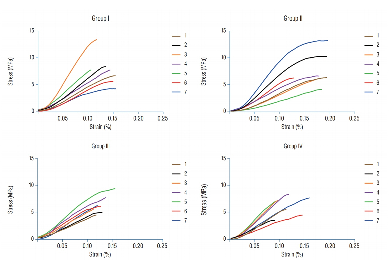
Fig. 5. This figure shows the curves resulting from the equations for the stress/strain ratio. Stress-strain curves for all groups (n=28) with a displacement rate of 10 mm/min.
Reference
-
References
1. Davignon KR, Dennehy KC. Update on postdural puncture headache. Int Anesthesiol Clin. 40:89–102. 2002.
Article2. Dufrane D, Marchal C, Cornu O, Raftopoulos C, Delloye C. Clinical application of a physically and chemically processed human substitute for dura mater. J Neurosurg. 98:1198–1202. 2003.
Article3. Famaey N, Verhoeven J, Jacobs S, Pettinari M, Meyns B. In situ evolution of the mechanical properties of stretchable and non-stretchable ePTFE vascular grafts and adjacent native vessels. Int J Artif Organs. 37:900–910. 2014.
Article4. Flaatten H, Felthaus J, Kuwelker M, Wisborg T. Postural post-dural puncture headache. A prospective randomised study and a meta-analysis comparing two different 0.40 mm O.D. (27 g) spinal needles. Acta Anaesthesiol Scand. 44:643–647. 2000.
Article5. Flaatten H, Thorsen T, Askeland B, Finne M, Rosland J, Hansen T, et al. Puncture technique and postural postdural puncture headache. A randomised, double-blind study comparing transverse and parallel puncture. Acta Anaesthesiol Scand. 42:1209–1214. 1998.
Article6. Kim M, Yoon H. Comparison of post-dural puncture headache and low back pain between 23 and 25 gauge Quincke spinal needles in patients over 60 years: randomized, double-blind controlled trial. Int J Nurs Stud. 48:1315–1322. 2011.
Article7. Lewis MC, Lafferty JP, Sacks MS, Pallares VS, TerRiet M. How much work is required to puncture dura with Tuohy needles? Br J Anaesth. 85:238–241. 2000.
Article8. Liu SS, McDonald SB. Current issues in spinal anesthesia. Anesthesiology. 94:888–906. 2001.
Article9. Longo S. Postdural puncture: implications and complications. Curr Opin Anaesthesiol. 12:271–275. 1999.
Article10. Lux EA, Althaus A. Is there a difference in postdural puncture headache after continuous spinal anesthesia with 28G microcatheters compared with punctures with 22G Quincke or Sprotte spinal needles? Local Reg Anesth. 7:63–67. 2014.
Article11. Lybecker H, Møller JT, May O, Nielsen HK. Incidence and prediction of postdural puncture headache. A prospective study of 1021 spinal anesthesias. Anesth Analg. 70:389–394. 1990.
Article12. Mahvash M, Dupont PE. Fast needle insertion to minimize tissue deformation and damage. IEEE Int Conf Robot Autom. 2009:3097–3102. 2009.
Article13. Matas SL. Why should we use atraumatic needles in lumbar puncture? Arq Neuropsiquiatr. 71:681–684. 2013.
Article14. McGarvey KA, Lee JM, Boughner DR. Mechanical suitability of glycerolpreserved human dura mater for construction of prosthetic cardiac valves. Biomaterials. 5:109–117. 1984.
Article15. Mihic DN. Postspinal headaches, needle surfaces and longitudinal orientation of the dural fibers. Results of a survey. Reg Anaesth. 9:54–56. 1986.16. Oh J, Liu K, Medina T, Kralick F, Noh HM. A novel microneedle array for the treatment of hydrocephalus. Microsyst Technol. 20:1169–1179. 2014.
Article17. Protasoni M, Sangiorgi S, Cividini A, Culuvaris GT, Tomei G, Dell’Orbo C, et al. The collagenic architecture of human dura mater. J Neurosurg. 114:1723–1730. 2011.
Article18. Reina MA, De Leon Casasola O, López A, De Andrés JA, Mora M, Fernández A. The origin of the spinal subdural space: ultrastructure findings. Anesth Analg. 94:991–995. table of contents. 2002.
Article19. Reina MA, López-García A, Dittmann M, de Andrés JA. Analysis of the external and internal surface of human dura mater with scanning electron microscopy. Rev Esp Anestesiol Reanim. 43:130–134. 1996.20. Schmittner MD, Urban N, Janke A, Weiss C, Bussen DG, Burmeister MA, et al. Influence of the pre-operative time in upright sitting position and the needle type on the incidence of post-dural puncture headache (PDPH) in patients receiving a spinal saddle block for anorectal surgery. Int J Colorectal Dis. 26:97–102. 2011.
Article21. Seeberger MD, Kaufmann M, Staender S, Schneider M, Scheidegger D. Repeated dural punctures increase the incidence of postdural puncture headache. Anesth Analg. 82:302–305. 1996.
Article22. van Gerwen DJ, Dankelman J, van den Dobbelsteen JJ. Needle-tissue interaction forces--a survey of experimental data. Med Eng Phys. 34:665–680. 2012.23. van Noort R, Black MM, Martin TR, Meanley S. A study of the uniaxial mechanical properties of human dura mater preserved in glycerol. Biomaterials. 2:41–45. 1981.
Article24. Wolfinbarger L Jr, Zhang YX, Adam BLT, Homsi D, Gates K, Sutherland V. Biomechanical aspects on rehydrated freeze-dried human allograft dura-mater tissues. J Appl Biomater. 5:265–270. 1994.
Article25. Yamada K, Miyamoto S, Nagata I, Kikuchi H, Ikada Y, Iwata H, et al. Development of a dural substitute from synthetic bioabsorbable polymers. J Neurosurg. 86:1012–1017. 1997.
Article26. Yamada K, Miyamoto S, Takayama M, Nagata I, Hashimoto N, Ikada Y, et al. Clinical application of a new bioabsorbable artificial dura mater. J Neurosurg. 96:731–735. 2002.
Article27. Zerris VA, James KS, Roberts JB, Bell E, Heilman CB. Repair of the dura mater with processed collagen devices. J Biomed Mater Res B Appl Biomater. 83:580–588. 2007.
Article
- Full Text Links
-
- Actions
-
Cited
- CITED
-
- Close
- Share
-
- Similar articles
-
- Comparison of Biomechanical Properties of Dura Mater Substitutes and Cranial Human Dura Mater : An In Vitro Study
- In vitro Fusion of the Posterior Frontal Calvarial Suture in the Mouse
- Dural Puncture by Introducer when Performing Spinal Anesthesia -A case report-
- Idiopathic Hypertrophic Cranial Pachymeningitis: Case Report
- Idiopathic Hypertrophic Pachymeningitis in the Craniocervical Junction
