J Periodontal Implant Sci.
2019 Oct;49(5):299-309. 10.5051/jpis.2019.49.5.299.
Cone-beam computed tomographic analysis of the alveolar ridge profile and virtual implant placement for the anterior maxilla
- Affiliations
-
- 1Department of Periodontology, Periodontal-Implant Clinical Research Institute, Kyung Hee University School of Dentistry, Seoul, Korea. shin.dmd@khu.ac.kr
- KMID: 2461020
- DOI: http://doi.org/10.5051/jpis.2019.49.5.299
Abstract
- PURPOSE
To analyze the ridge profile of the anterior maxilla using cone-beam computed tomography and to assess the clinical significance of the ridge profile by performing virtual implant placement.
METHODS
Thirty-two cone-beam computed tomography scans of anterior maxillae were included. For each tooth, a vertical line was made along the longitudinal axis, and 3 horizontal lines at 1-, 3-, and 5-mm levels below the labial bone crest were drawn perpendicularly to the vertical reference. At these levels, the thickness of the alveolar ridge (RT), and the labial (LT) and palatal bone plate (PT) were measured. Then, virtual implant placement using standard and tapered implants was performed. A generalized linear mixed model was used for statistical analysis.
RESULTS
The teeth were located labially based on the proportion of LT and PT with respect to RT. At the 1-mm level, the value of LT was between 1.0±0.4 mm for central incisors and 1.3±0.6 mm for canines. A large number of teeth had area(s) with less than 1-mm-thick labial bone between the 1- and 5-mm levels below the crest. The mean PT was generally thicker than the LT in all tooth types. The greatest mean value of labial concavity was observed for canines, compared to other tooth types. Men had a greater RT than did women, but had a comparable LT. Less apical fenestration was observed when tapered implants were used.
CONCLUSIONS
Most teeth in the anterior maxilla had a thin labial bone plate, with no significant difference between sexes. Tapered implants may be advantageous for the anterior maxilla.
MeSH Terms
Figure
-
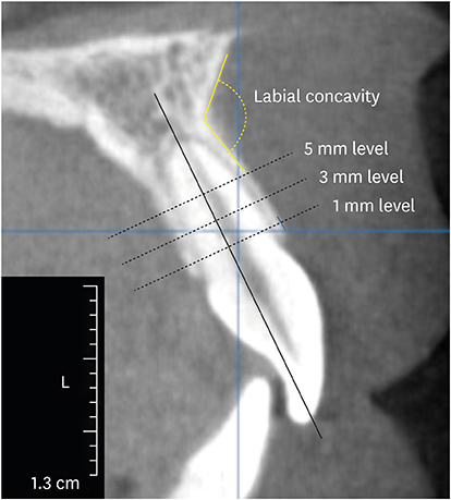
Figure 1 Measurements of cone-beam computed tomography scans.

Figure 2 Proportions of the teeth according to the LT. LT_1, LT_3, and LT_5 are LT at 1-, 3-, and 5-mm levels below the labial bone crest. LT: thickness of the labial bone plate.
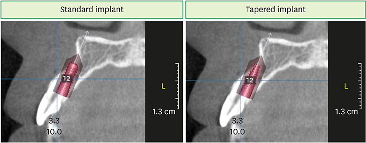
Figure 3 Virtual implant placement. Following implant placement (Ø3.3×10 mm), apical fenestration was observed in cases of standard implants, but not in cases of tapered implants.
Reference
-
1. Quirynen M, Herrera D, Teughels W, Sanz M. Implant therapy: 40 years of experience. Periodontol 2000. 2014; 66:7–12.2. Hämmerle CH, Chen ST, Wilson TG Jr. Consensus statements and recommended clinical procedures regarding the placement of implants in extraction sockets. Int J Oral Maxillofac Implants. 2004; 19 Suppl:26–28.3. Avila-Ortiz G, Elangovan S, Kramer KW, Blanchette D, Dawson DV. Effect of alveolar ridge preservation after tooth extraction: a systematic review and meta-analysis. J Dent Res. 2014; 93:950–958.
Article4. Cha JK, Song YW, Park SH, Jung RE, Jung UW, Thoma DS. Alveolar ridge preservation in the posterior maxilla reduces vertical dimensional change: a randomized controlled clinical trial. Clin Oral Implants Res. 2019; 30:515–523.
Article5. Lee JS, Cha JK, Kim CS. Alveolar ridge regeneration of damaged extraction sockets using deproteinized porcine versus bovine bone minerals: a randomized clinical trial. Clin Implant Dent Relat Res. 2018; 20:729–737.
Article6. Arora H, Ivanovski S. Immediate and early implant placement in single-tooth gaps in the anterior maxilla: a prospective study on ridge dimensional, clinical, and aesthetic changes. Clin Oral Implants Res. 2018; 29:1143–1154.
Article7. Covani U, Ricci M, Bozzolo G, Mangano F, Zini A, Barone A. Analysis of the pattern of the alveolar ridge remodelling following single tooth extraction. Clin Oral Implants Res. 2011; 22:820–825.
Article8. Schropp L, Wenzel A, Kostopoulos L, Karring T. Bone healing and soft tissue contour changes following single-tooth extraction: a clinical and radiographic 12-month prospective study. Int J Periodontics Restorative Dent. 2003; 23:313–323.9. Braut V, Bornstein MM, Belser U, Buser D. Thickness of the anterior maxillary facial bone wall-a retrospective radiographic study using cone beam computed tomography. Int J Periodontics Restorative Dent. 2011; 31:125–131.10. Bulyalert A, Pimkhaokham A. A novel classification of anterior alveolar arch forms and alveolar bone thickness: a cone-beam computed tomography study. Imaging Sci Dent. 2018; 48:191–199.
Article11. Januário AL, Duarte WR, Barriviera M, Mesti JC, Araújo MG, Lindhe J. Dimension of the facial bone wall in the anterior maxilla: a cone-beam computed tomography study. Clin Oral Implants Res. 2011; 22:1168–1171.
Article12. Zekry A, Wang R, Chau AC, Lang NP. Facial alveolar bone wall width - a cone-beam computed tomography study in Asians. Clin Oral Implants Res. 2014; 25:194–206.
Article13. Zhang W, Skrypczak A, Weltman R. Anterior maxilla alveolar ridge dimension and morphology measurement by cone beam computerized tomography (CBCT) for immediate implant treatment planning. BMC Oral Health. 2015; 15:65.
Article14. Araújo MG, Lindhe J. Dimensional ridge alterations following tooth extraction. An experimental study in the dog. J Clin Periodontol. 2005; 32:212–218.
Article15. Tao R, Meng M, Niu LN, Chen JH, Nico CF, Ma C. Investigation of sagittal root position in relation to the anterior maxillary alveolar bone: a cone-beam CT study in 300 cases with normal occlusion. Zhonghua Kou Qiang Yi Xue Za Zhi. 2017; 52:631–636.16. Buser D, Chappuis V, Belser UC, Chen S. Implant placement post extraction in esthetic single tooth sites: when immediate, when early, when late? Periodontol 2000. 2017; 73:84–102.
Article17. Kan JY, Rungcharassaeng K, Deflorian M, Weinstein T, Wang HL, Testori T. Immediate implant placement and provisionalization of maxillary anterior single implants. Periodontol 2000. 2018; 77:197–212.
Article18. Testori T, Weinstein T, Scutellà F, Wang HL, Zucchelli G. Implant placement in the esthetic area: criteria for positioning single and multiple implants. Periodontol 2000. 2018; 77:176–196.
Article19. Grunder U, Gracis S, Capelli M. Influence of the 3-D bone-to-implant relationship on esthetics. Int J Periodontics Restorative Dent. 2005; 25:113–119.20. Spray JR, Black CG, Morris HF, Ochi S. The influence of bone thickness on facial marginal bone response: stage 1 placement through stage 2 uncovering. Ann Periodontol. 2000; 5:119–128.
Article21. Kan JY, Roe P, Rungcharassaeng K, Patel RD, Waki T, Lozada JL, et al. Classification of sagittal root position in relation to the anterior maxillary osseous housing for immediate implant placement: a cone beam computed tomography study. Int J Oral Maxillofac Implants. 2011; 26:873–876.22. Lau SL, Chow J, Li W, Chow LK. Classification of maxillary central incisors-implications for immediate implant in the esthetic zone. J Oral Maxillofac Surg. 2011; 69:142–153.
Article23. Pascual A, Barallat L, Santos A, Levi P Jr, Vicario M, Nart J, et al. Comparison of periodontal biotypes between maxillary and mandibular anterior teeth: a clinical and radiographic study. Int J Periodontics Restorative Dent. 2017; 37:533–539.
Article24. Fuentes R, Flores T, Navarro P, Salamanca C, Beltrán V, Borie E. Assessment of buccal bone thickness of aesthetic maxillary region: a cone-beam computed tomography study. J Periodontal Implant Sci. 2015; 45:162–168.
Article25. El Nahass H, Naiem SN. Analysis of the dimensions of the labial bone wall in the anterior maxilla: a cone-beam computed tomography study. Clin Oral Implants Res. 2015; 26:e57–e61.
Article26. Lee SL, Kim HJ, Son MK, Chung CH. Anthropometric analysis of maxillary anterior buccal bone of Korean adults using cone-beam CT. J Adv Prosthodont. 2010; 2:92–96.
Article27. Schroeder HE. The periodontium. London: Springer;1986.28. Hubar JS. Quantification of the lamina dura. J Can Dent Assoc. 1993; 59:997–1000.29. Chen ST, Darby IB, Reynolds EC. A prospective clinical study of non-submerged immediate implants: clinical outcomes and esthetic results. Clin Oral Implants Res. 2007; 18:552–562.
Article30. Kan JY, Rungcharassaeng K, Sclar A, Lozada JL. Effects of the facial osseous defect morphology on gingival dynamics after immediate tooth replacement and guided bone regeneration: 1-year results. J Oral Maxillofac Surg. 2007; 65:13–19.
Article31. Chappuis V, Engel O, Shahim K, Reyes M, Katsaros C, Buser D. Soft tissue alterations in esthetic postextraction sites: a 3-dimensional analysis. J Dent Res. 2015; 94:187S–193S.
- Full Text Links
-
- Actions
-
Cited
- CITED
-
- Close
- Share
-
- Similar articles
-
- Horizontal alteration of anterior alveolar ridge after immediate implant placement: A retrospective cone beam computed tomography analysis
- Buccal cortical bone thickness on CBCT for mini-implant
- Invasion of the canalis sinuosus by dental implants: A report of 3 cases
- Alveolar Ridge Preservation in the Severely Damaged Sockets of the Anterior Maxilla Followed by Delayed Implant Placement
- Influence of the anterior arch shape and root position on root angulation in the maxillary esthetic area
