Imaging Sci Dent.
2019 Jun;49(2):123-130. 10.5624/isd.2019.49.2.123.
Influence of the anterior arch shape and root position on root angulation in the maxillary esthetic area
- Affiliations
-
- 1Esthetic Restorative and Implant Dentistry Program, Faculty of Dentistry, Chulalongkorn University, Bangkok, Thailand. atiphan.p@chula.ac.th
- 2Department of Prosthodontics, Faculty of Dentistry, Chulalongkorn University, Bangkok, Thailand.
- 3Department of Oral and Maxillofacial Surgery, Faculty of Dentistry, Chulalongkorn University, Bangkok, Thailand.
- KMID: 2450178
- DOI: http://doi.org/10.5624/isd.2019.49.2.123
Abstract
- PURPOSE
This study was conducted to characterize the relationship of the angulation between the tooth root axis and alveolar bone axis with anterior alveolar (AA) arch forms and sagittal root position (SRP) in the anterior esthetic region using cone-beam computed tomography (CBCT) images.
MATERIALS AND METHODS
CBCT images that met the inclusion and exclusion criteria were categorized using a recent classification of AA arch forms and a SRP classification. Then, the angulation of the root axis and the alveolar bone axis was measured using mid-sagittal CBCT images of each tooth. The relationships of the angulation with each AA arch form and SRP classification were evaluated using 1-way analysis of variance and a linear regression model.
RESULTS
Ninety-eight CBCT images were included in this study. SRP had a greater influence than the AA arch form on the angulation of the root axis and the alveolar bone axis (P<0.05). However, the combination of AA arch form and SRP was more predictive of the angulation of the root axis and the alveolar bone axis than either parameter individually.
CONCLUSION
The angulation of the root axis and alveolar bone axis demonstrated a relationship with the AA arch form and SRP in teeth in the anterior esthetic region. The influence of SRP was greater, but the combination of both parameters was more predictive of root-to-bone angulation than either parameter individually, implying that clinicians should account for both the AA arch form and SRP when planning implant placement procedures in this region.
MeSH Terms
Figure
-
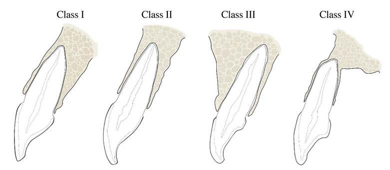
Fig. 1 The sagittal root position classification as reported by Kan et al.14

Fig. 2 The landmarks for angulation evaluation comprise the alveolar bone axis, tooth root axis, and the angle between the alveolar bone axis and the tooth root axis. A. Line A: alveolar bone axis, line 1: the buccal line; and line 2: the palatal line. B. Line B: the tooth root axis; line 3: the cervical line. C. C° is defined as the angle between the alveolar bone axis (line A) and the tooth root axis (line B).
Reference
-
1. Buser D, Martin W, Belser UC. Optimizing esthetics for implant restorations in the anterior maxilla: anatomic and surgical considerations. Int J Oral Maxillofac Implants. 2004; 19:Suppl. 43–61.2. Januário AL, Duarte WR, Barriviera M, Mesti JC, Araújo MG, Lindhe J. Dimension of the facial bone wall in the anterior maxilla: a cone-beam computed tomography study. Clin Oral Implants Res. 2011; 22:1168–1171.
Article3. Johnson K. A study of the dimensional changes occurring in the maxilla following tooth extraction. Aust Dent J. 1969; 14:241–244.
Article4. Misch CE. Anterior single-tooth replacement: surgical considerations. In : Misch CE, editor. Contemporary implant dentistry. 3rd ed. Canada: Mosby Elsevier;2008. p. 739–768.5. Moy PK, Pozzi A, Beumer J III. Surgical Considerations for the Esthetic zone. In : Beumer J, Faulkner RF, Shah K, Moy PK, editors. Fundamentals of implant dentistry: surgical principles. Hanover Park, IL: Quintessence Publishing;2016. p. 291–332.6. Jahangiri L, Devlin H, Ting K, Nishimura I. Current perspectives in residual ridge remodeling and its clinical implications: a review. J Prosthet Dent. 1998; 80:224–237.
Article7. Van der Weijden F, Dell'Acqua F, Slot DE. Alveolar bone dimensional changes of post-extraction sockets in humans: a systematic review. J Clin Periodontol. 2009; 36:1048–1058.
Article8. Ferrario VF, Sforza C, Miani A Jr, Tartaglia G. Mathematical definition of the shape of dental arches in human permanent healthy dentitions. Eur J Orthod. 1994; 16:287–294.
Article9. Preti G, Pera P, Bassi F. Prediction of the shape and size of the maxillary anterior arch in edentulous patients. J Oral Rehabil. 1986; 13:115–125.
Article10. Sagat G, Yalcin S, Gultekin BA, Mijiritsky E. Influence of arch shape and implant position on stress distribution around implants supporting fixed full-arch prosthesis in edentulous maxilla. Implant Dent. 2010; 19:498–508.
Article11. Pietrokovski J, Starinsky R, Arensburg B, Kaffe I. Morphologic characteristics of bony edentulous jaws. J Prosthodont. 2007; 16:141–147.
Article12. Suk KE, Park JH, Bayome M, Nam YO, Sameshima GT, Kook YA. Comparison between dental and basal arch forms in normal occlusion and Class III malocclusions utilizing cone-beam computed tomography. Korean J Orthod. 2013; 43:15–22.
Article13. Bulyalert A, Pimkhaokham A. A novel classification of anterior alveolar arch forms and alveolar bone thickness: a cone beam computed tomography study. Imaging Sci Dent. 2018; 48:191–199.14. Kan JY, Roe P, Rungcharassaeng K, Patel RD, Waki T, Lozada JL, et al. Classification of sagittal root position in relation to the anterior maxillary osseous housing for immediate implant placement: a cone beam computed tomography study. Int J Oral Maxillofac Implants. 2011; 26:873–876.15. Wang HM, Shen JW, Yu MF, Chen XY, Jiang QH, He FM. Analysis of facial bone wall dimensions and sagittal root position in the maxillary esthetic zone: a retrospective study using cone beam computed tomography. Int J Oral Maxillofac Implants. 2014; 29:1123–1129.
Article16. Zhang S, Shi X, Liu H. Angulations of anterior teeth with reference to the alveolar bone measured by CBCT in a Chinese population. Implant Dent. 2015; 24:397–401.
Article17. Masunaga M, Ueda H, Tanne K. Changes in the crown angulation and dental arch widths after nonextraction orthodontic treatment: model analysis of mild crowding with high canines. Open J Stomatol. 2012; 2:188–194.
Article18. Chan HL, Garaicoa-Pazmino C, Suarez F, Monje A, Benavides E, Oh TJ, et al. Incidence of implant buccal plate fenestration in the esthetic zone: a cone beam computed tomography study. Int J Oral Maxillofac Implants. 2014; 29:171–177.
Article19. Levine RA, Huynh-Ba G, Cochran DL. Soft tissue augmentation procedures for mucogingival defects in esthetic sites. Int J Oral Maxillofac Implants. 2014; 29:Suppl. 155–185.
Article20. Demircan S, Demircan E. Dental cone beam computed tomography analyses of the anterior maxillary bone thickness for immediate implant placement. Implant Dent. 2015; 24:664–668.
Article21. Lau SL, Chow J, Li W, Chow LK. Classification of maxillary central incisors-implications for immediate implant in the esthetic zone. J Oral Maxillofac Surg. 2011; 69:142–153.
Article22. Buser D, Chen ST, Weber HP, Belser UC. Early implant placement following single-tooth extraction in the esthetic zone: biologic rationale and surgical procedures. Int J Periodontics Restorative Dent. 2008; 28:441–451.23. Huynh-Ba G, Pjetursson BE, Sanz M, Cecchinato D, Ferrus J, Lindhe J, et al. Analysis of the socket bone wall dimensions in the upper maxilla in relation to immediate implant placement. Clin Oral Implants Res. 2010; 21:37–42.
Article24. Kim JH, Lee JG, Han DH, Kim HJ. Morphometric analysis of the anterior region of the maxillary bone for immediate implant placement using micro-CT. Clin Anat. 2011; 24:462–468.
Article25. Chung SH, Park YS, Chung SH, Shon WJ. Determination of implant position for immediate implant placement in maxillary central incisors using palatal soft tissue landmarks. Int J Oral Maxillofac Implants. 2014; 29:627–633.
Article26. Xu D, Wang Z, Sun L, Lin Z, Wan L, Li Y, et al. Classification of the root position of the maxillary central incisors and its clinical significance in immediate implant placement. Implant Dent. 2016; 25:520–524.
Article27. Jung YH, Cho BH, Hwang JJ. Analysis of the root position of the maxillary incisors in the alveolar bone using cone-beam computed tomography. Imaging Sci Dent. 2017; 47:181–187.
Article28. Garber DA, Belser UC. Restoration-driven implant placement with restoration-generated site development. Compend Contin Educ Dent. 1995; 16:796–804.29. Garber DA. The esthetic dental implant: letting restoration be the guide. J Am Dent Assoc. 1995; 126:319–325.
Article30. Grunder U, Gracis S, Capelli M. Influence of the 3-D bone-to-implant relationship on esthetics. Int J Periodontics Restorative Dent. 2005; 25:113–119.31. Cho YB, Moon SJ, Chung CH, Kim HJ. Resorption of labial bone in maxillary anterior implant. J Adv Prosthodont. 2011; 3:85–89.
Article32. Araujo MG, Linder E, Lindhe J. Bio-Oss collagen in the buccal gap at immediate implants: a 6-month study in the dog. Clin Oral Implants Res. 2011; 22:1–8.33. Mehta H, Shah S. Management of buccal gap and resorption of buccal plate in immediate implant placement: a clinical case report. J Int Oral Health. 2015; 7:Suppl 1. 72–75.
- Full Text Links
-
- Actions
-
Cited
- CITED
-
- Close
- Share
-
- Similar articles
-
- Assessment of Root and Root Canal Morphology of Human Primary Molars using CBCT
- A study on sagittal root position of maxillary anterior teeth in Korean
- Evaluation of Impacted Maxillary Canine Position Using Panoramic Radiographs and Cone-beam Computed Tomography
- Analysis of the root position and angulation of maxillary premolars in alveolar bone using cone-beam computed tomography
- Root and Canal Morphology of Maxillary Primary Molar using CBCT and 3D CT
