Primary Malignant Mesothelioma of the Peritoneum Mistaken for Peritoneal Tuberculosis due to Elevated Cancer Antigen 125
- Affiliations
-
- 1Division of Gastroenterology, Department of Internal Medicine, CHA Gumi Medical Center, CHA University School of Medicine, Gumi, Korea. zenus1@hanmail.net
- KMID: 2460989
- DOI: http://doi.org/10.4166/kjg.2019.74.4.232
Abstract
- A differential diagnosis of ascites is always challenging for physicians. Peritoneal tuberculosis is particularly difficult to distinguish from peritoneal carcinomatosis because of the similarities in clinical manifestations and laboratory results. Although the definitive diagnostic method for ascites is to take a biopsy of the involved tissues through laparoscopy or laparotomy, there are many limitations in performing biopsies in clinical practice. For this reason, physicians have attempted to find surrogate markers that can substitute for a biopsy as a confirmative diagnostic method for ascites. CA 125, which is known as a tumor marker for gynecological malignancies, has been reported to be a biochemical indicator for peritoneal tuberculosis. On the other hand, the sensitivity of serum CA 125 is low, and CA 125 may be elevated due to other benign or malignant conditions. This paper reports the case of a 66-year-old male who had a moderate amount of ascites and complained of dyspepsia and a febrile sensation. His abdominal CT scans revealed a conglomerated mass, diffuse omental infiltration, and peritoneal wall thickening. Initially, peritoneal tuberculosis was suspected due to the clinical symptoms, CT findings, and high serum CA 125 levels, but non-specific malignant cells were detected on cytology of the ascitic fluid. Finally, he was diagnosed with primary malignant peritoneal mesothelioma after undergoing a laparoscopic biopsy.
MeSH Terms
Figure
-
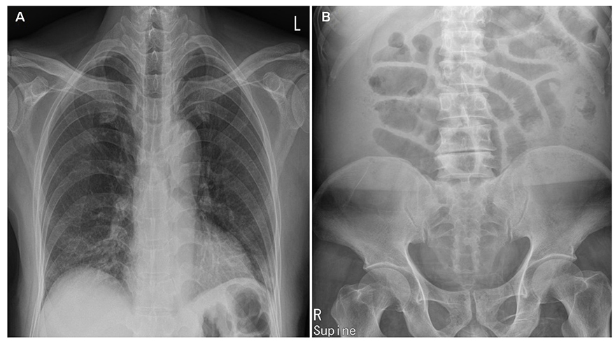
Fig. 1 Chest X-ray and simple abdomen scan images. (A) The chest X-ray shows increased bronchovascular markings in the right lower lung filed, but no active lung disease and pleural effusion. (B) The centralized bowel gases are noted due to massive ascites in the simple abdomen erect view.

Fig. 2 Contrast-enhanced computed tomography images of the abdomen show a 4.4×5.5 cm conglomerated mass (yellow arrows) among the portal vein, pancreas neck, and liver. Diffuse omental infiltration, a moderate amount of ascites and peritoneal thickening are also revealed.
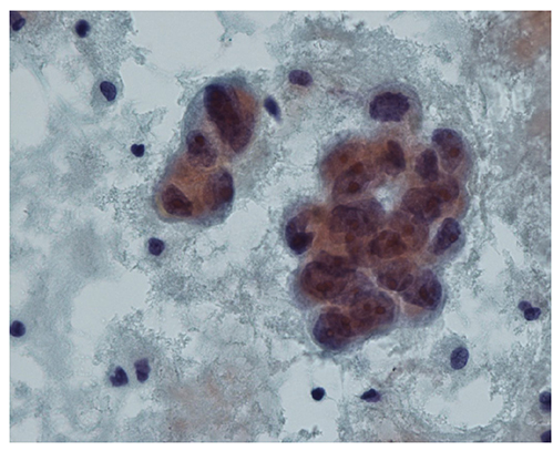
Fig. 3 Aspiration of ascites shows suspicious malignant cells, which were finally revealed as atypical mesothelial cells (ThinPrep 2000 System; Cytyc Corp., Marlborough, MA, USA) (Papanicolaou stain, ×400).
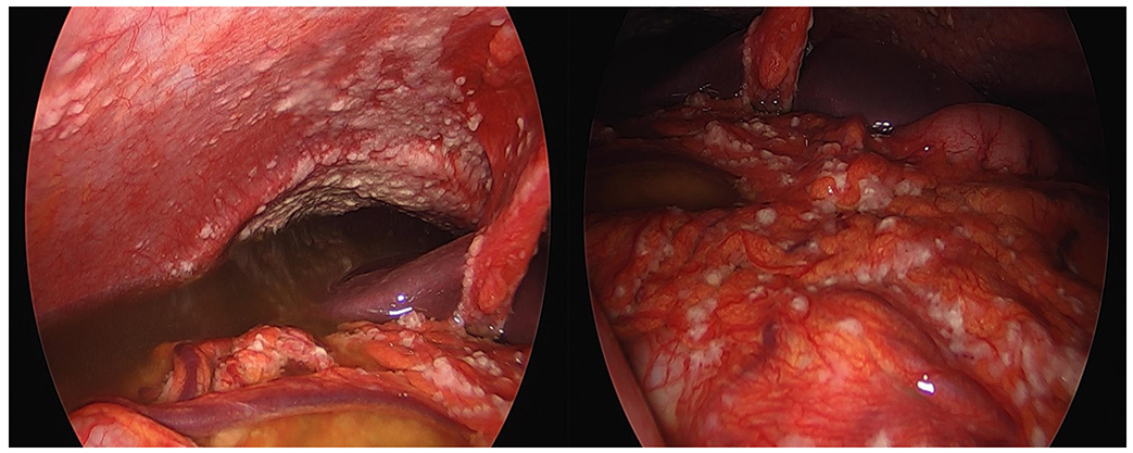
Fig. 4 Gross photographs during laparoscopic biopsy reveal a large amount of ascites and multiple whitish patch nodules within the whole peritoneum, omentum, and bowel walls.
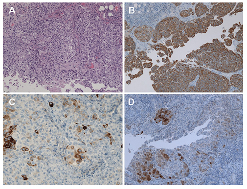
Fig. 5 Histology specimen shows malignant mesothelial cells, epitheloid type (A: H&E, ×100). Immunohistochemical staining of the specimens is reactive for cytokeratin and cytokeratin-7 and focally positive for calretinin, confirming the diagnosis of malignant peritoneal mesothelioma (B: cytokeratin, ×100; C: cytokeratin-7, ×200; D: calretinin, ×100).
Reference
-
1. Lee JY. Diagnosis and treatment of extrapulmonary tuberculosis. Tuberc Respir Dis (Seoul). 2015; 78:47–55.
Article2. Kim JH, Yim JJ. Achievements in and challenges of tuberculosis control in South Korea. Emerg Infect Dis. 2015; 21:1913–1920.
Article3. Mas MR, Cömert B, Sağlamkaya U, et al. CA-125; a new marker for diagnosis and follow-up of patients with tuberculous peritonitis. Dig Liver Dis. 2000; 32:595–597.
Article4. Rasheed S, Zinicola R, Watson D, Bajwa A, McDonald PJ. Intra-abdominal and gastrointestinal tuberculosis. Colorectal Dis. 2007; 9:773–783.
Article5. Kocaman O. Understanding tuberculous peritonitis: a difficult task to overcome. Turk J Gastroenterol. 2014; 25:79–80.
Article6. Shih CA, Ho SP, Tsay FW, Lai KH, Hsu PI. Diffuse malignant peritoneal mesothelioma. Kaohsiung J Med Sci. 2013; 29:642–645.
Article7. Torrejón Reyes PN, Frisancho O, Gómez A, Yábar A. Malignant peritoneal mesothelioma. Rev Gastroenterol Peru. 2010; 30:82–87.8. Melero M, Lloveras J, Waisman H, Elsner B, Baldessari E. Malignant peritoneal mesothelioma. An infrequent cause of prolonged fever syndrome and leucocytosis in a young adult. Medicina (B Aires). 1995; 55:48–50.9. Yin WJ, Zheng GQ, Chen YF, et al. CT differentiation of malignant peritoneal mesothelioma and tuberculous peritonitis. Radiol Med. 2016; 121:253–260.
Article10. Silberstein LB, Rosenthal AN, Coppack SW, Noonan K, Jacobs IJ. Ascites and a raised serum CA 125--confusing combination. J R Soc Med. 2001; 94:581–582.
Article11. Thakur V, Mukherjee U, Kumar K. Elevated serum cancer antigen 125 levels in advanced abdominal tuberculosis. Med Oncol. 2001; 18:289–291.
Article12. Seo BS, Hwang IK, Ra JE, Kim YS. A patient with tuberculous peritonitis with very high serum CA 125. BMJ Case Rep. 2012; 2012:bcr2012006382.
Article13. Kaya M, Kaplan MA, Isikdogan A, Celik Y. Differentiation of tuberculous peritonitis from peritonitis carcinomatosa without surgical intervention. Saudi J Gastroenterol. 2011; 17:312–317.
Article14. Choi CH, Kim CJ, Lee YY, et al. Peritoneal tuberculosis: a retrospective review of 20 cases and comparison with primary peritoneal carcinoma. Int J Gynecol Cancer. 2010; 20:798–803.
Article15. Bae SY, Lee JH, Park JY, et al. Clinical significance of serum CA-125 in Korean females with ascites. Yonsei Med J. 2013; 54:1241–1247.
Article16. Kebapci M, Vardareli E, Adapinar B, Acikalin M. CT findings and serum ca 125 levels in malignant peritoneal mesothelioma: report of 11 new cases and review of the literature. Eur Radiol. 2003; 13:2620–2626.
Article17. Gube M, Taeger D, Weber DG, et al. Performance of biomarkers SMRP, CA125, and CYFRA 21-1 as potential tumor markers for malignant mesothelioma and lung cancer in a cohort of workers formerly exposed to asbestos. Arch Toxicol. 2011; 85:185–192.
Article18. Hollevoet K, Nackaerts K, Gosselin R, et al. Soluble mesothelin, megakaryocyte potentiating factor, and osteopontin as markers of patient response and outcome in mesothelioma. J Thorac Oncol. 2011; 6:1930–1937.
Article19. Rump A, Morikawa Y, Tanaka M, et al. Binding of ovarian cancer antigen CA125/MUC16 to mesothelin mediates cell adhesion. J Biol Chem. 2004; 279:9190–9198.
Article20. Kaneko O, Gong L, Zhang J, et al. A binding domain on mesothelin for CA125/MUC16. J Biol Chem. 2009; 284:3739–3749.
Article
- Full Text Links
-
- Actions
-
Cited
- CITED
-
- Close
- Share
-
- Similar articles
-
- A Case of Papillary Serous Carcinoma of the Peritoneum
- Two Cases of Primary Carcinoma of the Peritoneum
- Two cases of malignant mesothelioma of the peritoneum and pericardium
- Malignant peritoneal mesothelioma with nonspecific history: a case report
- A Case of Diffuse Malignant Mesothelioma of the Peritoneum
