Investig Magn Reson Imaging.
2019 Sep;23(3):241-250. 10.13104/imri.2019.23.3.241.
An MRI-Based Quantification for Correlation of Imaging Biomarker and Clinical Performance in Chronic Phase of Carbon Monoxide Poisoning
- Affiliations
-
- 1Department of Radiology, Soonchunhyang University Bucheon Hospital, Bucheon, Korea.
- 2Department of Radiology, Soonchunhyang University Seoul Hospital, Seoul, Korea. stpark@schmc.ac.kr
- 3Department of Radiology, Hallym University Dongtan Sacred Heart Hospital, Dongtan, Korea.
- 4Department of Radiology, Soonchunhyang University Cheonan Hospital, Cheonan, Korea.
- KMID: 2459878
- DOI: http://doi.org/10.13104/imri.2019.23.3.241
Abstract
- PURPOSE
The purpose of this study was to determine the relation between quantitative magnetic resonance imaging biomarkers, and clinical performances in chronic phase of carbon monoxide intoxication.
MATERIALS AND METHODS
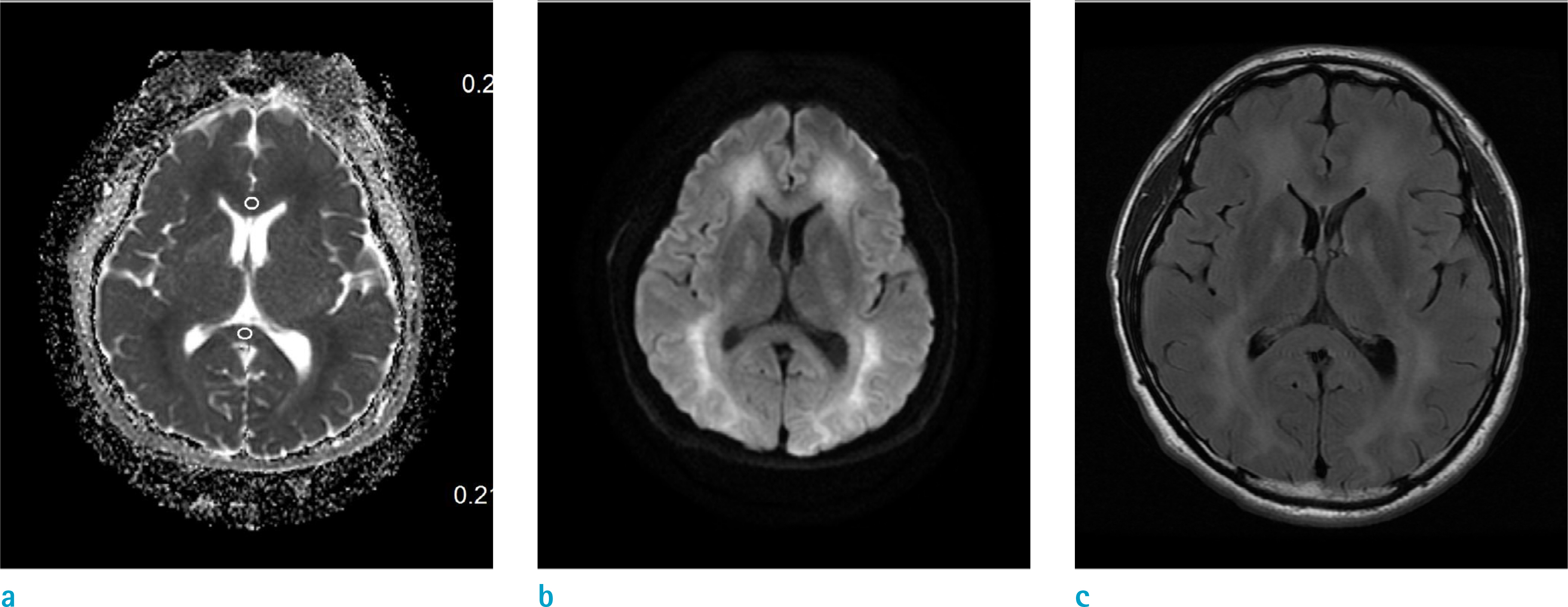
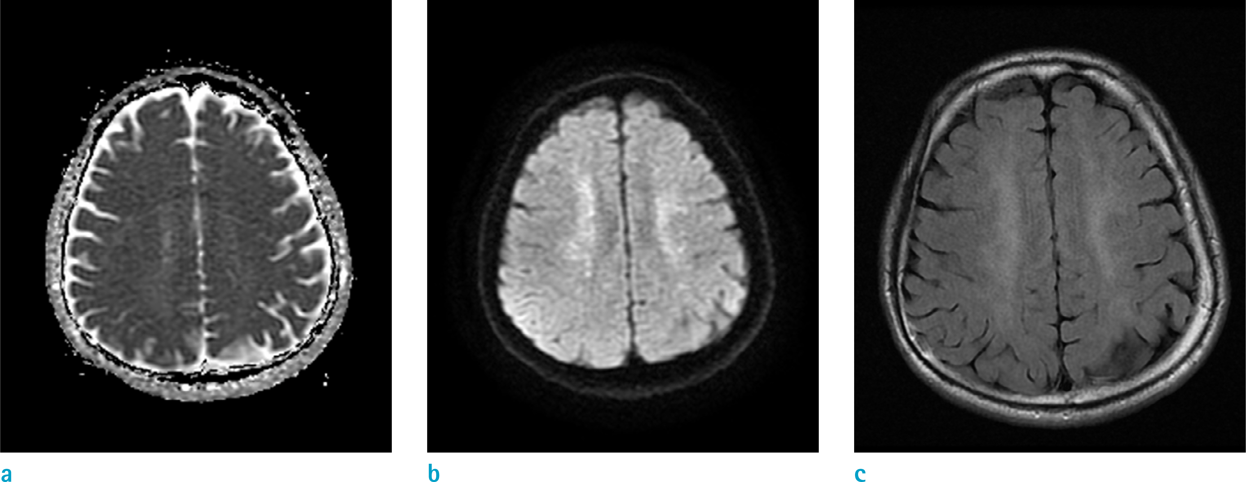
Eighteen magnetic resonance scans and cognitive evaluations were performed, on patients with carbon monoxide intoxication in chronic phase. Apparent diffusion coefficient (ADC) ratios of affected versus unaffected centrum semiovale, and corpus callosum were obtained. Signal intensity (SI) ratios between affected centrum semiovale, and normal pons in T2-FLAIR (fluid-attenuated inversion recovery) images were obtained. The Mini-Mental State Exam, and clinical outcome scores were assessed. Correlation coefficients were calculated, between MRI and clinical markers. Patients were further classified into poor-outcome and good-outcome groups based on clinical performance, and imaging parameters were compared. T2-SI ratio of centrum semiovale was compared, with that of 18 sex-matched and age-matched controls.
RESULTS
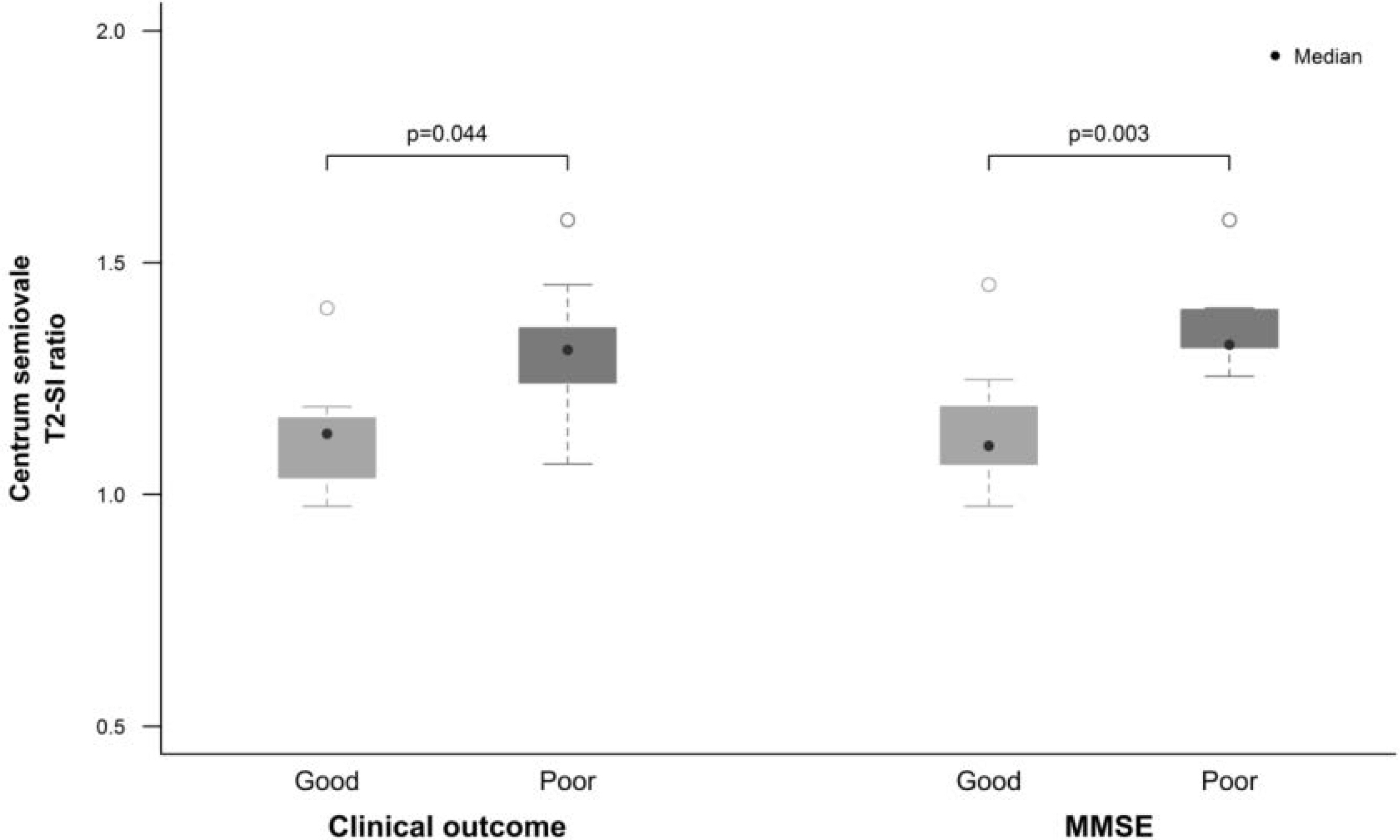
T2-SI ratio of centrum semiovale was significantly higher in the poor-outcome group, than that in the good-outcome group and was strongly inversely correlated, with results from the Mini-Mental State Exam. ADC ratios of centrum semiovale were significantly lower in the poor outcome group than in the good outcome group, and were moderately correlated with the Mini-Mental State Exam score.
CONCLUSION
A higher T2-SI and a lower ratio of ADC values in the centrum semiovale, may indicate presence of more severe white matter injury and clinical impairment. T2-SI ratio and ADC values in the centrum semiovale, are useful quantitative imaging biomarkers for correlation with clinical performance in individuals with carbon monoxide intoxication.
Keyword
MeSH Terms
Figure
Reference
-
References
21. Pracyk JB, Stolp BW, Fife CE, Gray L, Piantadosi CA. Brain computerized tomography after hyperbaric oxygen therapy for carbon monoxide poisoning. Undersea Hyperb Med. 1995; 22:1–7.22. Sener RN. Acute carbon monoxide poisoning: diffusion MR imaging findings. AJNR Am J Neuroradiol. 2003; 24:1475–1477.23. Murata T, Kimura H, Kado H, et al. Neuronal damage in the interval form of CO poisoning determined by serial diffusion weighted magnetic resonance imaging plus 1H-magnetic resonance spectroscopy. J Neurol Neurosurg Psychiatry. 2001; 71:250–253.
Article24. Pierpaoli C, Righini A, Linfante I, Tao-Cheng JH, Alger JR, Di Chiro G. Histopathologic correlates of abnormal water diffusion in cerebral ischemia: diffusion-weighted MR imaging and light and electron microscopic study. Radiology. 1993; 189:439–448.
Article25. Beppu T, Fujiwara S, Nishimoto H, et al. Fractional anisotropy in the centrum semiovale as a quantitative indicator of cerebral white matter damage in the subacute phase in patients with carbon monoxide poisoning: correlation with the concentration of myelin basic protein in cerebrospinal fluid. J Neurol. 2012; 259:1698–1705.
Article26. Fujiwara S, Beppu T, Nishimoto H, et al. Detecting damaged regions of cerebral white matter in the subacute phase after carbon monoxide poisoning using voxel-based analysis with diffusion tensor imaging. Neuroradiology. 2012; 54:681–689.
Article27. Pandya DN, Karol EA, Heilbronn D. The topographical distribution of interhemispheric projections in the corpus callosum of the rhesus monkey. Brain Res. 1971; 32:31–43.
Article
- Full Text Links
- Actions
-
Cited
- CITED
-
- Close
- Share
- Similar articles
-
- Cerebral Infarction Followed by Hemorrhagic Transformation Accompanied by Carbon Monoxide Poisoning
- Pulmonary edema in acute carbon monoxide poisoning
- A Clinical Observation of Acute Carbon Monoxide Poisoning
- Chorea as a Clinical Manifestation of Delayed Neurologic Sequelae in Carbon Monoxide Poisoning: Case Report
- Influence of Acute and Chronic Carbon Monoxide Poisoning on Reproductive Organs of the White Rats: Enzymological Study 2. Lactate Dehydrogenase Activity in the Prostate


