The Role of Double Inversion Recovery Imaging in Acute Ischemic Stroke
- Affiliations
-
- 1Department of Radiology, Kyung Hee University Hospital at Gangdong, College of Medicine, Kyung Hee University, Seoul, Korea. ghjahng@gmail.com
- KMID: 2459875
- DOI: http://doi.org/10.13104/imri.2019.23.3.210
Abstract
- PURPOSE
The purpose of this study was to investigate if double inversion recovery (DIR) imaging can have a role in the evaluation of brain ischemia, compared with diffusion-weighted imaging (DWI) and fluid-attenuated inversion recovery (FLAIR) imaging.
MATERIALS AND METHODS
Sixty-seven patients within 48 hours of onset, underwent MRI scans with FLAIR, DWI with b-value of 0 (B0) and 1000 s/mm², and DIR sequences. Patients were categorized into four groups: within three hours, three to six hours, six to 24 hours, and 24 to 48 hours after onset. Lesion-to-normal ratio (LNR) value was calculated and compared among all sequences within each group, by the Friedman test and conducted among all groups, for each sequence by the Kruskal-Wallis test. In qualitative assessment, signal intensity changes of DIR, B0, and FLAIR based on similarity with DWI and image quality of each sequence, were graded on a 3-point scale, respectively. Scores for detectability of lesions were compared by the McNemar's test.
RESULTS
LNR values from DWI were higher than DIR, but not statistically significant in all groups (P > 0.05). LNR values of DIR were significantly higher than FLAIR within 24 hours of onset (P < 0.05). LNR values were significantly different between, before, and after six hours onset time for DIR (P = 0.016), B0 (P = 0.008), and FLAIR (P = 0.018) but not for DWI (P = 0.051). Qualitative analysis demonstrated that detectability of DIR was higher, compared to that of FLAIR within 4.5 hours and six hours of onset (P < 0.05). Also, the DWI quality score was lower than that of DIR, particularly relative to infratentorial lesions.
CONCLUSION
DIR provides higher detectability of hyperacute brain ischemia than B0 and FLAIR, and does not suffer from susceptibility artifact, unlike DWI. So, DIR can be used to replace evaluation of the FLAIR-DWI mismatch.
Keyword
MeSH Terms
Figure
-
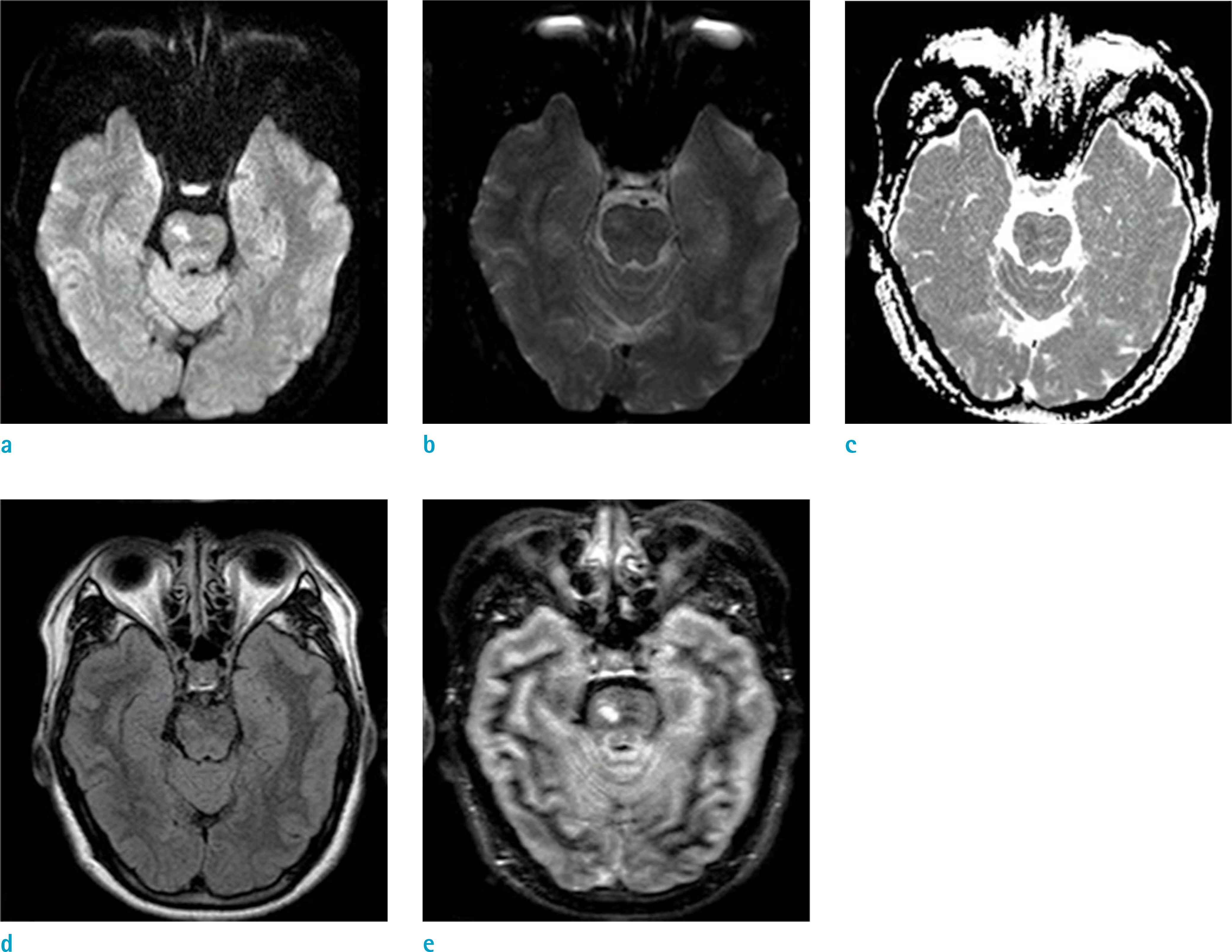
Fig. 1. Representative magnetic resonance imaging (MRI) images scanned 24 hours after symptom onset, in the case of 50 years-old female with infarction in the right pons. Diffusion-weighted imaging (DWI) ambiguously shows the ischemic lesion in the right pons, but double inversion recovery (DIR) depicts the lesion more clearly than the other sequences. From left to right, images are from (a) DWI, (b) DWI with b-value of 0s/mm2 (B0), (c) Apparent diffusion coefficient (ADC), (d) Fluid-attenuated inversion recovery (FLAIR) and (e) DIR. Note that the LNR values of each sequence are as follow: DWI = 43.5, DIR = 22.7, FLAIR = 9.4, and B0 = 5.8.
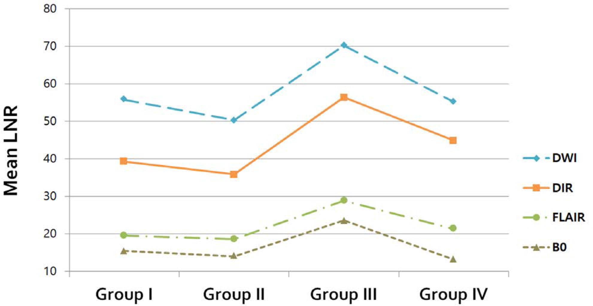
Fig. 2. Representative magnetic resonance imaging (MRI) images, collected three hours after symptom onset of 71 years-old female. There is ischemic infarction, in the right middle cerebral artery (MCA) territory. Diffusion-weighted imaging (DWI) depicts the lesion more clearly than the other sequences; Double inversion recovery (DIR) also depicts the lesion better than fluid-attenuated inversion recovery (FLAIR) and DWI with b-value of 0s/mm2 (B0) sequences. From left to right, images are from (a) B0, (b) DWI, (c) Apparent diffusion coefficient (ADC), (d) DIR and (e) FLAIR. Note that LNR values of each sequence are as follow: DWI = 32.8, DIR = 60.3, FLAIR = 10.2, and B0 = 22.2.
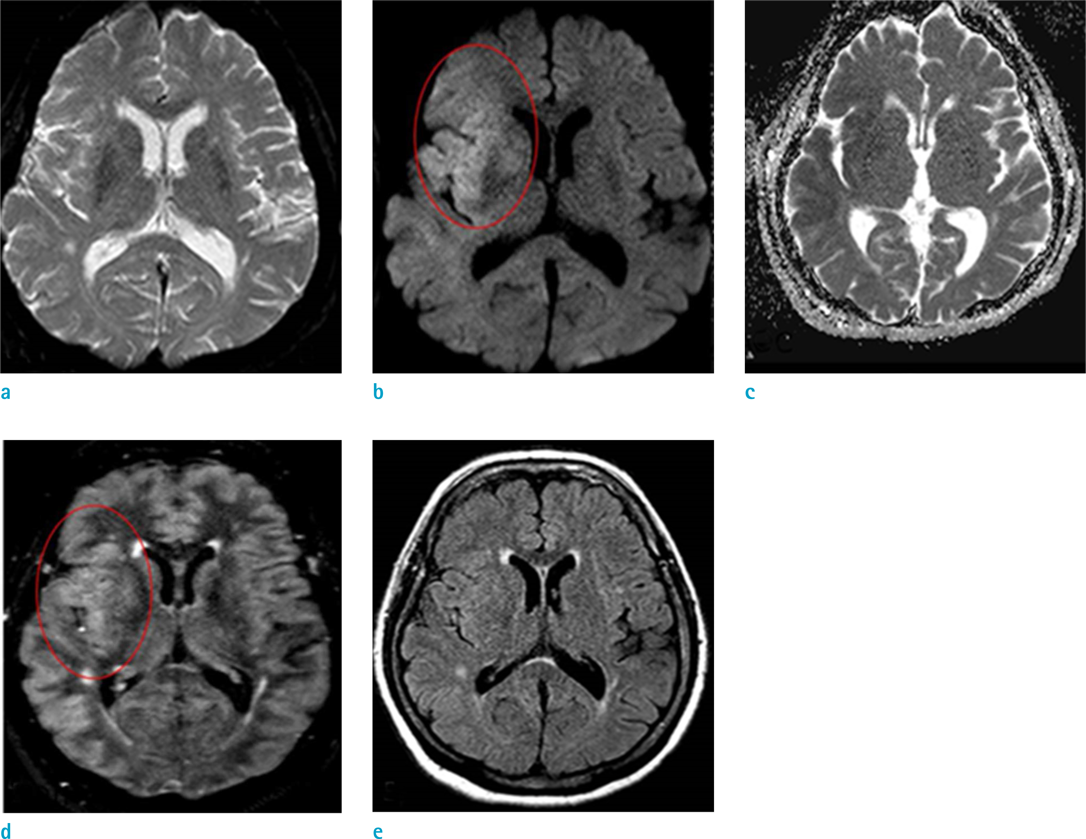
Fig. 3. Graphs of mean lesion-to-normal ratio (LNR) values, of each sequence within all groups (Group I through Group IV).
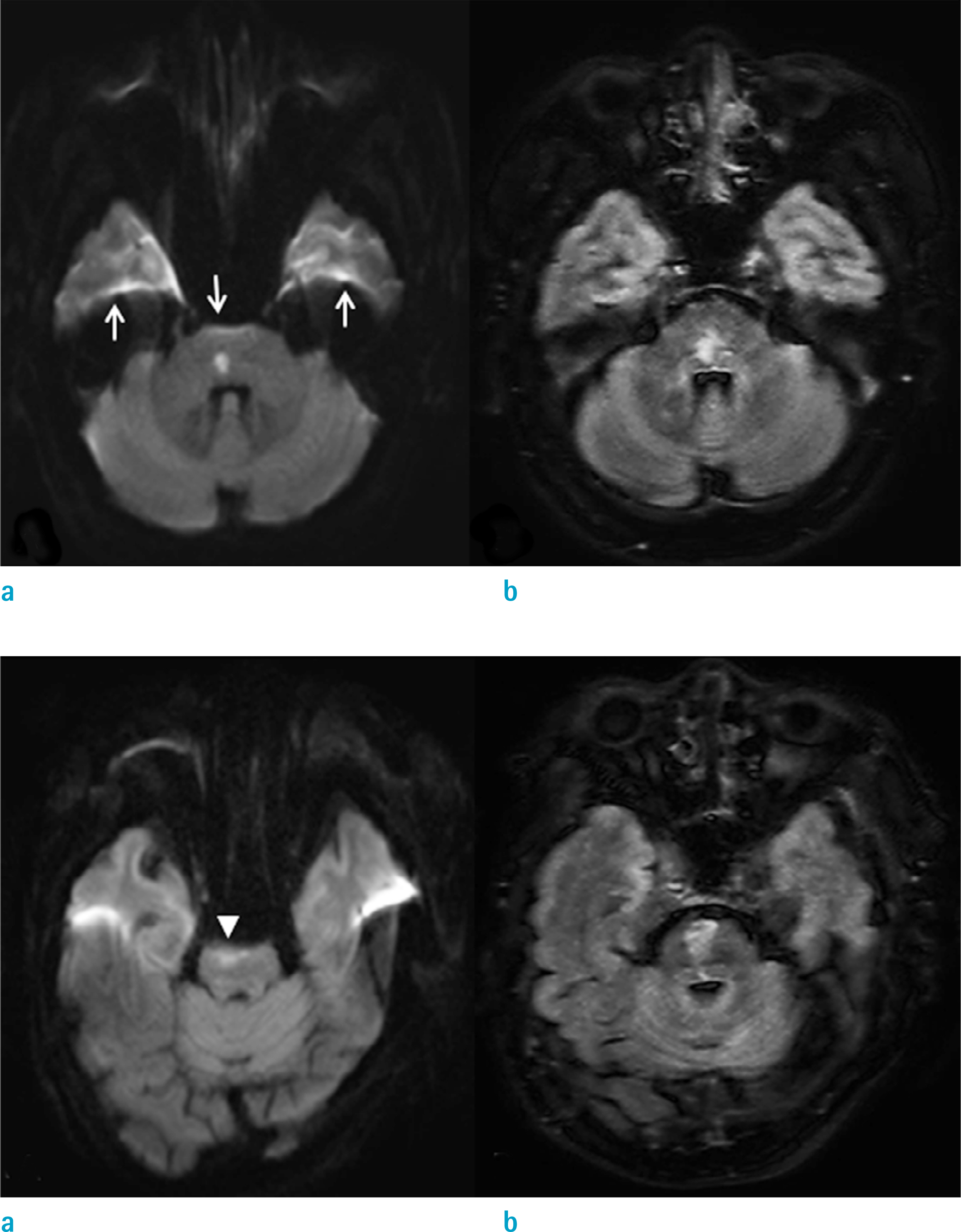
Fig. 4. Examples of two patients with infratentorial infarctions, for qualitative assessment of image quality in each sequence. Top row: An age 50 male who underwent magnetic resonance imaging (MRI) scan at 20 hours after left ataxia. A typical susceptibility artifact (white arrows) is visible on (a) diffusion-weighted imaging (DWI), without affecting diagnosis of a small (< 2 cm) hyperintense lesion in the right pons (Score 2). In (b) double inversion recovery (DIR), a lesion is identified at a location corresponding to that of DWI, without anatomic distortion (Score 3). Bottom row: Images of an age 81 female presenting left side weakness and speech disturbance, were obtained within 48 hours after symptom onset. (a) DWI shows considerable geometric distortion by susceptibility artifact, leading to underestimation of right paramedian pontine infarction (arrowhead) (Score 1). Also, itself may be mistaken for an artifact. In contrast, (b) DIR shows definite high signal intensity lesion in the right pons (Score 3).
Reference
-
References
1. Benveniste H, Hedlund LW, Johnson GA. Mechanism of detection of acute cerebral ischemia in rats by diffusion-weighted magnetic resonance microscopy. Stroke. 1992; 23:746–754.
Article2. Brant-Zawadzki M, Atkinson D, Detrick M, Bradley WG, Scidmore G. Fluid-attenuated inversion recovery (FLAIR) for assessment of cerebral infarction. Initial clinical experience in 50 patients. Stroke. 1996; 27:1187–1191.3. Gauvrit JY, Leclerc X, Girot M, et al. Fluid-attenuated inversion recovery (FLAIR) sequences for the assessment of acute stroke: inter observer and inter technique reproducibility. J Neurol. 2006; 253:631–635.4. Le Bihan D, Poupon C, Amadon A, Lethimonnier F. Artifacts and pitfalls in diffusion MRI. J Magn Reson Imaging. 2006; 24:478–488.
Article5. Lutsep HL, Albers GW, DeCrespigny A, Kamat GN, Marks MP, Moseley ME. Clinical utility of diffusion-weighted magnetic resonance imaging in the assessment of ischemic stroke. Ann Neurol. 1997; 41:574–580.
Article6. Sorensen AG, Buonanno FS, Gonzalez RG, et al. Hyperacute stroke: evaluation with combined multisection diffusion-weighted and hemodynamically weighted echoplanar MR imaging. Radiology. 1996; 199:391–401.
Article7. Perkins CJ, Kahya E, Roque CT, Roche PE, Newman GC. Fluid-attenuated inversion recovery and diffusion- and perfusion-weighted MRI abnormalities in 117 consecutive patients with stroke symptoms. Stroke. 2001; 32:2774–2781.
Article8. Alexander JA, Sheppard S, Davis PC, Salverda P. Adult cerebrovascular disease: role of modified rapid fluid-attenuated inversion-recovery sequences. AJNR Am J Neuroradiol. 1996; 17:1507–1513.9. Reidel MA, Stippich C, Heiland S, Storch-Hagenlocher B, Jansen O, Hahnel S. Differentiation of multiple sclerosis plaques, subacute cerebral ischaemic infarcts, focal vasogenic oedema and lesions of subcortical arteriosclerotic encephalopathy using magnetisation transfer measurements. Neuroradiology. 2003; 45:289–294.
Article10. Ricci PE, Burdette JH, Elster AD, Reboussin DM. A comparison of fast spin-echo, fluid-attenuated inversion-recovery, and diffusion-weighted MR imaging in the first 10 days after cerebral infarction. AJNR Am J Neuroradiol. 1999; 20:1535–1542.11. Thomalla G, Cheng B, Ebinger M, et al. DWI-FLAIR mismatch for the identification of patients with acute ischaemic stroke within 4.5 h of symptom onset (PRE-FLAIR): a multicentre observational study. Lancet Neurol. 2011; 10:978–986.
Article12. Aoki J, Kimura K, Iguchi Y, Shibazaki K, Sakai K, Iwanaga T. FLAIR can estimate the onset time in acute ischemic stroke patients. J Neurol Sci. 2010; 293:39–44.
Article13. Wattjes MP, Lutterbey GG, Gieseke J, et al. Double inversion recovery brain imaging at 3T: diagnostic value in the detection of multiple sclerosis lesions. AJNR Am J Neuroradiol. 2007; 28:54–59.14. Jahng GH, Stables L, Ebel A, et al. Sensitive and fast T1 mapping based on two inversion recovery images and a reference image. Med Phys. 2005; 32:1524–1528.
Article15. Geurts JJ, Pouwels PJ, Uitdehaag BM, Polman CH, Barkhof F, Castelijns JA. Intracortical lesions in multiple sclerosis: improved detection with 3D double inversion-recovery MR imaging. Radiology. 2005; 236:254–260.
Article16. Rugg-Gunn FJ, Boulby PA, Symms MR, Barker GJ, Duncan JS. Imaging the neocortex in epilepsy with double inversion recovery imaging. Neuroimage. 2006; 31:39–50.
Article17. Calabrese M, De Stefano N, Atzori M, et al. Detection of cortical inflammatory lesions by double inversion recovery magnetic resonance imaging in patients with multiple sclerosis. Arch Neurol. 2007; 64:1416–1422.
Article18. Nelson F, Poonawalla AH, Hou P, Huang F, Wolinsky JS, Narayana PA. Improved identification of intracortical lesions in multiple sclerosis with phase-sensitive inversion recovery in combination with fast double inversion recovery MR imaging. AJNR Am J Neuroradiol. 2007; 28:1645–1649.
Article19. Jahng GH, Lee DK, Lee JM, Rhee HY, Ryu CW. Double inversion recovery imaging improves the evaluation of gray matter volume losses in patients with Alzheimer's disease and mild cognitive impairment. Brain Imaging Behav. 2016; 10:1015–1028.
Article20. Bedell BJ, Narayana PA. Implementation and evaluation of a new pulse sequence for rapid acquisition of double inversion recovery images for simultaneous suppression of white matter and CSF. J Magn Reson Imaging. 1998; 8:544–547.
Article21. Turetschek K, Wunderbaldinger P, Bankier AA, et al. Double inversion recovery imaging of the brain: initial experience and comparison with fluid attenuated inversion recovery imaging. Magn Reson Imaging. 1998; 16:127–135.
Article22. Redpath TW, Smith FW. Technical note: use of a double inversion recovery pulse sequence to image selectively grey or white brain matter. Br J Radiol. 1994; 67:1258–1263.23. Boulby PA, Symms MR, Barker GJ. Optimized interleaved whole-brain 3D double inversion recovery (DIR) sequence for imaging the neocortex. Magn Reson Med. 2004; 51:1181–1186.
Article24. Hacke W, Warach S. Diffusion-weighted MRI as an evolving standard of care in acute stroke. Neurology. 2000; 54:1548–1549.
Article25. Benameur K, Bykowski JL, Luby M, Warach S, Latour LL. Higher prevalence of cortical lesions observed in patients with acute stroke using high-resolution diffusion-weighted imaging. AJNR Am J Neuroradiol. 2006; 27:1987–1989.26. Cotton F, Rambaud L, Hermier M. Dual inversion recovery MRI helps identifying cortical tubers in tuberous sclerosis. Epilepsia. 2006; 47:1072–1073.
Article27. Noguchi K, Ogawa T, Inugami A, et al. Acute subarachnoid hemorrhage: MR imaging with fluid-attenuated inversion recovery pulse sequences. Radiology. 1995; 196:773–777.
Article
- Full Text Links
-
- Actions
-
Cited
- CITED
-
- Close
- Share
-
- Similar articles
-
- Fast MRI in Acute Ischemic Stroke: Applications of MRI Acceleration Techniques for MR-Based Comprehensive Stroke Imaging
- Hyperintense Vessels on FLAIR MRI in Patients With Acute Middle Cerebral Artery Infarction Revealed Pial Collateral on Cerebral Angiography
- Usefulness of Diffusion - Weighted Imaging in Acute and Subacute Ischemic Stroke: Comparison with Fast Spin-Echo T2-Weighted Imaging and Fluid Attenuated Inversion Recovery Imaging
- Pneumococcal meningitis complicated by otomastoiditis and pneumocephalus confounding an acute ischemic stroke diagnosis
- Is There a Difference in the Effect of Thrombolytic Therapy according to the Presence of Diffusion-Weighted Imaging (DWI)-Fluid Attenuated Inversion Recovery (FLAIR) Mismatching in Patients with Acute Ischemic Stroke?
