Korean J Radiol.
2019 Oct;20(10):1454-1461. 10.3348/kjr.2018.0841.
Ultrasound-Guided Core Needle Biopsy Techniques for Intermediate or Low Suspicion Thyroid Nodules: Which Method is Effective for Diagnosis?
- Affiliations
-
- 1Department of Radiology and Center for Imaging Science, Thyroid Center, Samsung Medical Center, Sungkyunkwan University School of Medicine, Seoul, Korea. helena35@hanmail.net
- 2Department of Pathology, Thyroid Center, Samsung Medical Center, Sungkyunkwan University School of Medicine, Seoul, Korea.
- KMID: 2459164
- DOI: http://doi.org/10.3348/kjr.2018.0841
Abstract
OBJECTIVE
To retrospectively compare the diagnostic performances of two different ultrasound (US)-guided core needle biopsy (CNB) techniques for intermediate or low suspicion thyroid nodules.
MATERIALS AND METHODS
Between August 2015 and December 2016, two different biopsy techniques were alternatively applied for 248 consecutive thyroid nodules, of which, 140 intermediate or low suspicion thyroid nodules were included in this study. In the first technique, two specimens included nodular tissue, nodular margin, and surrounding normal parenchyma (i.e., marginal target). In the second technique, two specimens were obtained from two different target areas, one for the marginal target and another for the intranodular target. The diagnostic performances of the two techniques to predict neoplasm and malignancy were compared.
RESULTS
CNB was performed on 80 intermediate or low suspicion nodules (57.1%) using the first technique and on 60 (42.9%) using the second technique. The accuracy of the first technique for predicting neoplasm or malignancy was significantly higher than that of the second technique (100% vs. 93.3%, p = 0.032 for predicting neoplasm; 88.8% vs. 75.0%, p = 0.033 for predicting malignancy). The negative predictive value of the first technique for predicting malignancy was also significantly higher than that of the second technique (87.5% vs. 72.7%, p = 0.035).
CONCLUSION
For intermediate or low suspicion thyroid nodules, US-guided CNB to obtain two specimens with marginal targets is more effective for diagnosing neoplasm or malignancy than is CNB for respective marginal and intranodular targets.
MeSH Terms
Figure
-
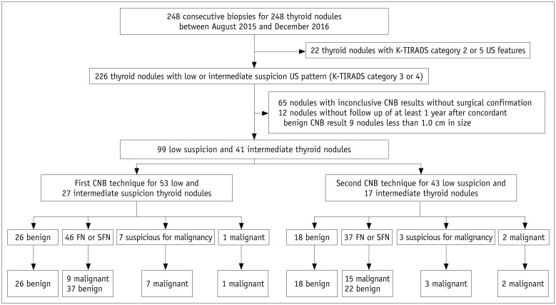
Fig. 1 Study flow and outcomes of study population.Numbers represent number of thyroid nodules. CNB = core needle biopsy, FN or SFN = follicular neoplasm or suspicious for follicular neoplasm, K-TIRADS = Korean Thyroid Imaging Reporting and Data System, US = ultrasound
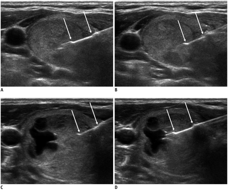
Fig. 2 Two CNB techniques.In first biopsy technique (A, B), two specimens included nodular tissue, nodular margin, and surrounding normal parenchyma (i.e., margin target). In second technique (C, D), two specimens were obtained from two different target areas, one from marginal target (C) and another from intranodular target (D). Arrows indicate specimen notch of biopsy needle.
Reference
-
1. Yeon JS, Baek JH, Lim HK, Ha EJ, Kim JK, Song DE, et al. Thyroid nodules with initially nondiagnostic cytologic results: the role of core-needle biopsy. Radiology. 2013; 268:274–280. PMID: 23525204.
Article2. Choi YJ, Baek JH, Suh CH, Shim WH, Jeong B, Kim JK, et al. Core-needle biopsy versus repeat fine-needle aspiration for thyroid nodules initially read as atypia/follicular lesion of undetermined significance. Head Neck. 2017; 39:361–369. PMID: 27704650.
Article3. Yoon RG, Baek JH, Lee JH, Choi YJ, Hong MJ, Song DE, et al. Diagnosis of thyroid follicular neoplasm: fine-needle aspiration versus core-needle biopsy. Thyroid. 2014; 24:1612–1617. PMID: 25089716.
Article4. Lee HY, Baek JH, Ha EJ, Park JW, Lee JH, Song DE, et al. Malignant-looking thyroid nodules with size reduction: core needle biopsy results. Ultrasonography. 2016; 35:327–334. PMID: 27184652.
Article5. Na DG, Baek JH, Jung SL, Kim JH, Sung JY, Kim KS, et al. Korean Society of Thyroid Radiology (KSThR) and Korean Society of Radiology. Core needle biopsy of the thyroid: 2016 consensus statement and recommendations from Korean Society of Thyroid Radiology. Korean J Radiol. 2017; 18:217–237. PMID: 28096731.
Article6. Suh CH, Baek JH, Lee JH, Choi YJ, Kim JK, Sung TY, et al. The role of core-needle biopsy as a first-line diagnostic tool for initially detected thyroid nodules. Thyroid. 2016; 26:395–403. PMID: 26651390.
Article7. Kim HC, Kim YJ, Han HY, Yi JM, Baek JH, Park SY, et al. First-line use of core needle biopsy for high-yield preliminary diagnosis of thyroid nodules. AJNR Am J Neuroradiol. 2017; 38:357–363. PMID: 27932508.
Article8. Suh CH, Baek JH, Choi YJ, Kim TY, Sung TY, Song DE, et al. Efficacy and safety of core-needle biopsy in initially detected thyroid nodules via propensity score analysis. Sci Rep. 2017; 7:8242. PMID: 28811482.
Article9. Chung SR, Suh CH, Baek JH, Choi YJ, Lee JH. The role of core needle biopsy in the diagnosis of initially detected thyroid nodules: a systematic review and meta-analysis. Eur Radiol. 2018; 28:4909–4918. PMID: 29789911.
Article10. Han S, Shin JH, Hahn SY, Oh YL. Modified core biopsy technique to increase diagnostic yields for well-circumscribed indeterminate thyroid nodules: a retrospective analysis. AJNR Am J Neuroradiol. 2016; 37:1155–1159. PMID: 26846928.
Article11. Hahn SY, Shin JH, Oh YL. What is the ideal core number for ultrasonography-guided thyroid biopsy of cytologically inconclusive nodules? AJNR Am J Neuroradiol. 2017; 38:777–781. PMID: 28154123.
Article12. Ahn S, Jung S, Kim JY, Shin JH, Hahn SY, Oh YL. Evaluation of modified core-needle biopsy in the diagnosis of thyroid nodules. Korean J Radiol. 2018; 19:656–664. PMID: 29962871.
Article13. Kim JH, Na DG, Lee H. Ultrasonographic echogenicity and histopathologic correlation of thyroid nodules in core needle biopsy specimens. Korean J Radiol. 2018; 19:673–681. PMID: 29962873.
Article14. Shin JH, Baek JH, Chung J, Ha EJ, Kim JH, Lee YH, et al. Korean Society of Thyroid Radiology (KSThR) and Korean Society of Radiology. Ultrasonography diagnosis and imaging-based management of thyroid nodules: revised Korean Society of Thyroid Radiology consensus statement and recommendations. Korean J Radiol. 2016; 17:370–395. PMID: 27134526.
Article15. Jung CK, Min HS, Park HJ, Song DE, Kim JH, Park SY, et al. Pathology reporting of thyroidcore needle biopsy: a proposal of the Korean Endocrine Pathology Thyroid Core Needle Biopsy Study Group. J Pathol Transl Med. 2015; 49:288–299. PMID: 26081825.16. American Thyroid Association (ATA) Guidelines Taskforce on Thyroid Nodules and Differentiated Thyroid Cancer. Cooper DS, Doherty GM, Haugen BR, Kloos RT, Lee SL, Mandel SJ, et al. Revised American Thyroid Association management guidelines for patients with thyroid nodules and differentiated thyroid cancer. Thyroid. 2009; 19:1167–1214. PMID: 19860577.
Article17. Cibas ES, Ali SZ. NCI Thyroid FNA State of the Science Conference. The Bethesda System For Reporting Thyroid Cytopathology. Am J Clin Pathol. 2009; 132:658–665. PMID: 19846805.
Article18. Baloch ZW, Livolsi VA. Follicular-patterned lesions of the thyroid: the bane of the pathologist. Am J Clin Pathol. 2002; 117:143–150. PMID: 11789719.19. Schreiner AM, Yang GC. Adenomatoid nodules are the main cause for discrepant histology in 234 thyroid fine-needle aspirates reported as follicular neoplasm. Diagn Cytopathol. 2012; 40:375–379. PMID: 22508673.
Article20. Hong MJ, Na DG, Baek JH, Sung JY, Kim JH. Cytology-ultrasonography risk-stratification scoring system based on fine-needle aspiration cytology and the Korean-Thyroid Imaging Reporting and Data System. Thyroid. 2017; 27:953–959. PMID: 28463597.
Article21. Rago T, Di Coscio G, Basolo F, Scutari M, Elisei R, Berti P, et al. Combined clinical, thyroid ultrasound and cytological features help to predict thyroid malignancy in follicular and Hupsilonrthle cell thyroid lesions: results from a series of 505 consecutive patients. Clin Endocrinol (Oxf). 2007; 66:13–20. PMID: 17201796.22. Jeh SK, Jung SL, Kim BS, Lee YS. Evaluating the degree of conformity of papillary carcinoma and follicular carcinoma to the reported ultrasonographic findings of malignant thyroid tumor. Korean J Radiol. 2007; 8:192–197. PMID: 17554185.
Article23. Sillery JC, Reading CC, Charboneau JW, Henrichsen TL, Hay ID, Mandrekar JN. Thyroid follicular carcinoma: sonographic features of 50 cases. AJR Am J Roentgenol. 2010; 194:44–54. PMID: 20028904.
Article24. Hahn SY, Shin JH, Oh YL, Kim TH, Lim Y, Choi JS. Role of ultrasound in predicting tumor invasiveness in follicular variant of papillary thyroid carcinoma. Thyroid. 2017; 27:1177–1184. PMID: 28699414.
Article25. Park KT, Ahn SH, Mo JH, Park YJ, Park DJ, Choi SI, et al. Role of core needle biopsy and ultrasonographic finding in management of indeterminate thyroid nodules. Head Neck. 2011; 33:160–165. PMID: 20848434.
Article26. Renshaw AA, Pinnar N. Comparison of thyroid fine-needle aspiration and core needle biopsy. Am J Clin Pathol. 2007; 128:370–374. PMID: 17709309.
Article27. Screaton NJ, Berman LH, Grant JW. US-guided core-needle biopsy of the thyroid gland. Radiology. 2003; 226:827–832. PMID: 12601219.
Article28. Ha EJ, Baek JH, Lee JH, Kim JK, Choi YJ, Sung TY, et al. Complications following US-guided core-needle biopsy for thyroid lesions: a retrospective study of 6,169 consecutive patients with 6,687 thyroid nodules. Eur Radiol. 2017; 27:1186–1194. PMID: 27311538.
Article29. Ha EJ, Suh CH, Baek JH. Complications following ultrasound-guided core needle biopsy of thyroid nodules: a systematic review and meta-analysis. Eur Radiol. 2018; 28:3848–3860. PMID: 29589112.
Article30. Nyquist GG, Tom WD, Mui S. Automatic core needle biopsy: a diagnostic option for head and neck masses. Arch Otolaryngol Head Neck Surg. 2008; 134:184–189. PMID: 18283162.31. Na DG, Kim JH, Sung JY, Baek JH, Jung KC, Lee H, et al. Core-needle biopsy is more useful than repeat fine-needle aspiration in thyroid nodules read as nondiagnostic or atypia of undetermined significance by the Bethesda system for reporting thyroid cytopathology. Thyroid. 2012; 22:468–475. PMID: 22304417.
Article
- Full Text Links
-
- Actions
-
Cited
- CITED
-
- Close
- Share
-
- Similar articles
-
- Effectiveness and Limitations of Core Needle Biopsy in the Diagnosis of Thyroid Nodules: Review of Current Literature
- Follow-up of benign thyroid nodules confirmed by ultrasound-guided core needle biopsy after inconclusive cytology on fine-needle aspiration biopsy
- Thyroid Nodules with Nondiagnostic FNA Results: Role of Core Needle Biopsy
- The Utility of US-Guided Core-Needle Biopsy in the Diagnosis of Thyroid Nodules
- Two Cases of Thyroid Hematoma Developing after a Core Needle Biopsy
