Imaging of Thoracic Wall Abnormalities
- Affiliations
-
- 1Department of Diagnostic Radiology, McGill University Health Centre, Montreal General Hospital, Montreal, Canada. alexandre.semionov@mail.mcgill.ca
- 2Department of Diagnostic Radiology, McGill University Health Centre, Montreal, Canada.
- 3Department of Radiology, King Abdulaziz University, Faculty of Medicine, Jeddah, Saudi Arabia.
- 4Department of Radiology, Jewish General Hospital, Montreal, Canada.
- KMID: 2459163
- DOI: http://doi.org/10.3348/kjr.2019.0181
Abstract
- Identification of certain abnormalities of the chest wall can be extremely helpful in correctly diagnosing a number of syndromic conditions and systemic diseases. Additionally, chest wall abnormalities may sometimes constitute diagnoses by themselves. In the present pictorial essay, we review a number of such conditions and provide illustrative cases that were retrospectively identified from our clinical imaging database. These include pentalogy of Cantrell, Klippel-Feil syndrome, cleidocranial dysplasia, Poland syndrome, osteopetrosis, neurofibromatosis type 1, Marfan syndrome, Gardner syndrome, systemic sclerosis, relapsing polychondritis, polymyositis/dermatomyositis, ankylosing spondylitis, hyperparathyroidism, rickets, sickle cell anemia, thalassemia, tuberculosis, septic arthritis of the sternoclavicular joint, elastofibroma dorsi, and sternal dehiscence.
Keyword
MeSH Terms
-
Anemia, Sickle Cell
Arthritis, Infectious
Cleidocranial Dysplasia
Diagnosis
Gardner Syndrome
Hyperparathyroidism
Klippel-Feil Syndrome
Marfan Syndrome
Neurofibromatosis 1
Osteopetrosis
Pentalogy of Cantrell
Poland Syndrome
Polychondritis, Relapsing
Retrospective Studies
Rickets
Scleroderma, Systemic
Spondylitis, Ankylosing
Sternoclavicular Joint
Thalassemia
Thoracic Wall*
Tuberculosis
Figure
-

Fig. 1 Pentalogy of Cantrell.Chest CT scan of 25-year-old male with Pentalogy of Cantrell, status post remote Fontan procedure for complex congenital heart abnormality, shows absence of sternum resulting in partial herniation of right ventricle (arrowheads). Note extracardiac Fontan conduit (arrow), multiple mediastinal collateral vessels, and bilateral pleural effusions.
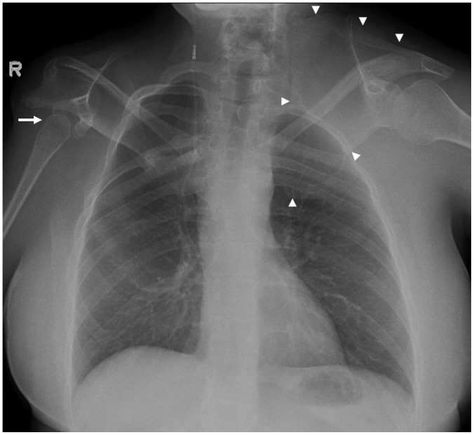
Fig. 2 Klippel-Feil syndrome.Chest radiograph of 39-year-old female with Klippel-Feil syndrome shows hypoplastic right humeral head (arrow), dysmorphic right scapula and glenoid, high-riding left scapula-Sprengel deformity (arrowheads), and multiple upper rib deformities. There is right ventriculo-peritoneal shunt.
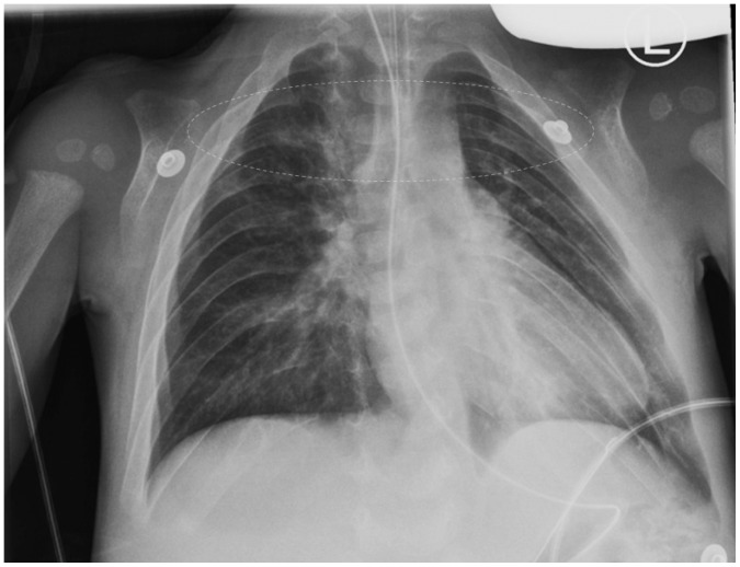
Fig. 3 Cleidocranial dysplasia.Chest radiograph of 3-year-old male with cleidocranial dysplasia shows absence of both clavicles and slanting of ribs. Expected location of clavicles is outlined by dashed line.
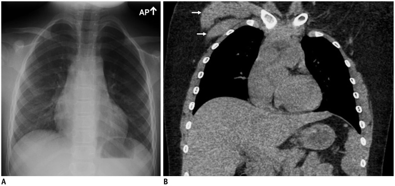
Fig. 4 Poland syndrome.10-year-old female with Poland syndrome.A. Chest radiograph demonstrates hyperlucent left hemithorax. B. Chest CT with coronal reformation shows absence of left pectoral muscles. Note normal right pectoral muscles (arrows).
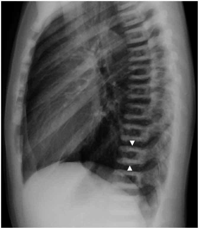
Fig. 5 Osteopetrosis.Lateral chest radiograph of 14-year-old male with osteopetrosis shows abnormally increased density of all bones and characteristic “sandwich vertebrae” (arrowheads).

Fig. 6 Neurofibromatosis type 1.Chest CT scan of 50-year-old female with neurofibromatosis type 1 demonstrates multiple cutaneous neurofibromas (arrowheads).

Fig. 7 Neurofibromatosis type 1.Chest CT with coronal reformation in 16-year-old female with neurofibromatosis type 1 demonstrates multiple rib deformities and bilateral intercostal plexiform neurofibromas (arrows).
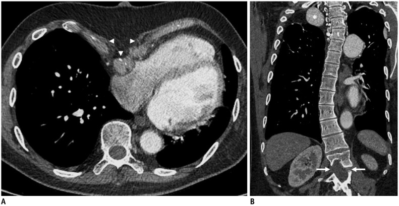
Fig. 8 Marfan syndrome.Axial CT angiogram (A) and CT with coronal reformation (B) of 25-year-old male with Marfan syndrome show pectus excavatum (arrowheads), scoliosis, upper lumbar dural ectasia (arrows), and aneurysm of right subclavian artery (asterisk).

Fig. 9 Gardner syndrome.Chest CT scan of 35-year-old female patient with Gardner syndrome demonstrates bilateral anterior chest wall lesions (arrows), which are biopsy-proven desmoid tumors.
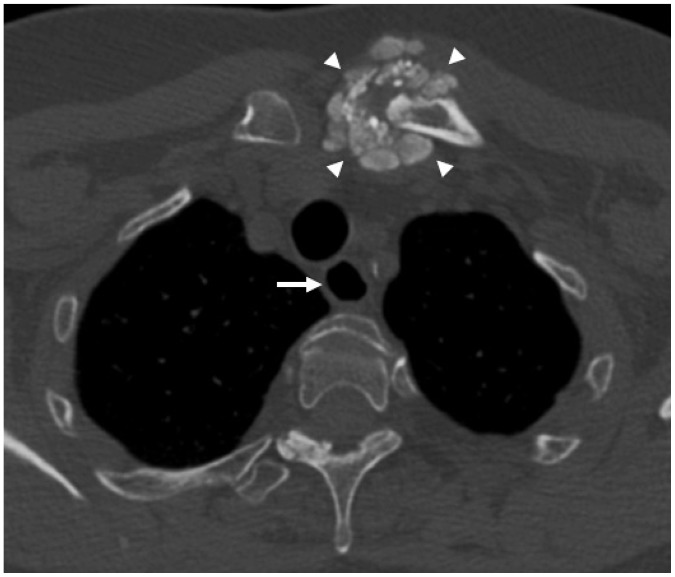
Fig. 10 Systemic sclerosis.Chest CT scan of 54-year-old female with systemic sclerosis shows exuberant amorphous calcifications around left sternoclavicular joint (arrowheads) and patulous esophagus (arrow).
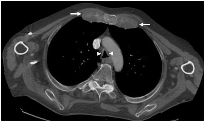
Fig. 11 Relapsing polychondritis.Chest CT scan of 77-year-old male with relapsing polychondritis demonstrates marked thickening of costal cartilages (arrows), as well as thickening of tracheal wall (arrowheads) with sparing of posterior membrane and tracheal luminal narrowing.
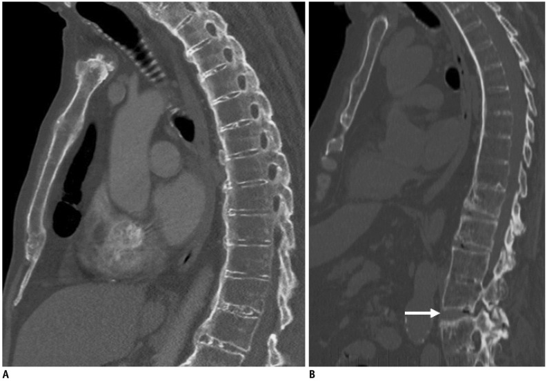
Fig. 12 Ankylosing spondylitis.A. CT with sagittal reformation in 76-year-old male with ankylosing spondylitis shows diffuse ankylosis of thoracolumbar spine. B. Chest CT with sagittal reformation in different patient with ankylosing spondylitis shows acute Chance fracture in upper lumbar spine (arrow).
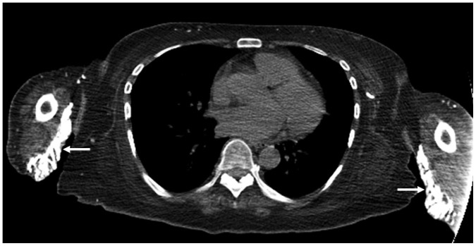
Fig. 13 Dermatomyositis.Chest CT scan of 51-year-old female with severe dermatomyositis shows diffuse atrophy of chest wall muscles and extensive subcutaneous calcifications in upper arms (arrows).

Fig. 14 Hyperparathyroidism.Frontal (A) and lateral (B) chest radiographs of 31-year-old male with secondary hyperparathyroidism due to chronic renal failure show resorption of distal clavicles (arrowheads) and “rugger-jersey” spine.

Fig. 15 Rickets.Frontal chest radiograph of 3-month-old premature-born female with rickets demonstrates healing fractures of left posterior 6th and 7th ribs (arrows), nodular widening of bilateral costochondral junctions-rachitic rosary (arrowheads) and fraying of proximal humeral metaphyses (dashed outlines).

Fig. 16 Sickle cell anemia.Frontal (A) and lateral (B) chest radiographs of 39-year-old female with sickle cell anemia demonstrate H-shaped vertebrae (arrowheads) and left humeral head subchondral sclerosis (arrow), consistent with avascular necrosis.
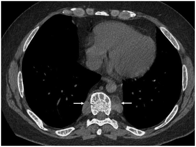
Fig. 17 Beta thalassemia.Chest CT scan of 44-year-old male with beta thalassemia intermedia shows diffuse expansion of osseous medullary spaces, coarsened trabeculations of ribs and vertebrae, and paravertebral extramedullary hematopoiesis (arrows).

Fig. 18 Beta thalassemia.Sagittal T1-weighted MRI of spine in 9-year-old male with beta thalassemia major shows diffuse abnormally low signal intensity of bone marrow related to active red marrow and iron deposition from multiple blood transfusions. Note that bone marrow (asterisk) is of significantly lower intensity than intervertebral disks (arrowhead).
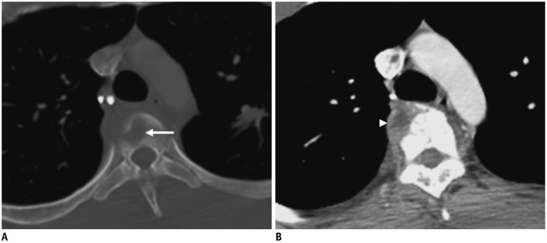
Fig. 19 Tuberculosis.Chest CT in bone (A) and mediastinal (B) windows of 23-year-old male with active post-primary pulmonary tuberculosis and spondylitis demonstrates irregular lytic lesion involving superior endplate of dorsal vertebra (arrow) and associated perivertebral abscess (arrowhead).
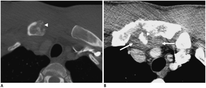
Fig. 20 Septic arthritis of sternoclavicular joint.Chest CT in bone (A) and mediastinal (B) windows of 41-year-old male, who developed septic arthritis of right sternoclavicular joint several weeks following penetrating injury to chest, demonstrates erosive lesions in right clavicular head and manubrium (arrowheads) consistent with osteomyelitis, and adjacent phlegmonous collection (arrows).

Fig. 21 Elastofibroma dorsi.Chest CT scan of 81-year-old female shows bilateral soft-tissue masses between scapulae and rib cage (arrows), consistent with bilateral elastofibroma dorsi.
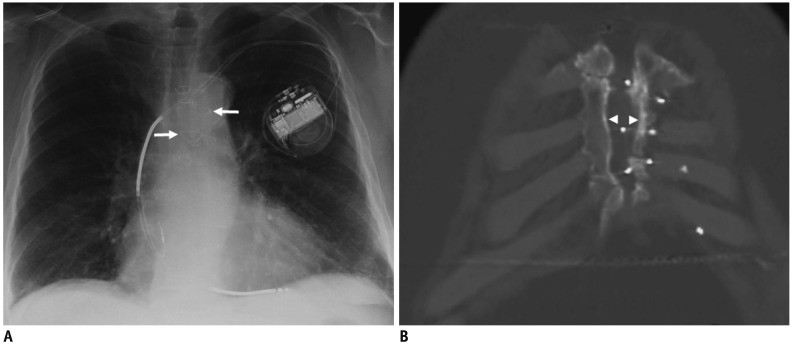
Fig. 22 Sternal dehiscence.72-year-old male with chronic sternal dehiscence following remote median sternotomy for aorto-coronary bypass surgery.A. Chest radiograph shows fractures and misalignment of many sternal wires (arrows). B. Chest CT with coronal reformation shows separation of sternotomy edges (arrowheads).
Reference
-
1. van Hoorn JH, Moonen RM, Huysentruyt CJ, van Heurn LW, Offermans JP, Mulder AL. Pentalogy of Cantrell: two patients and a review to determine prognostic factors for optimal approach. Eur J Pediatr. 2008; 167:29–35. PMID: 17674044.
Article2. Samartzis D, Herman J, Lubicky JP, Shen FH. Sprengel's deformity in Klippel-Feil syndrome. Spine (Phila Pa 1976). 2007; 32:E512–E516. PMID: 17700431.
Article3. Paul SA, Simon SS, Karthik AK, Chacko RK, Savitha S. A review of clinical and radiological features of cleidocranial dysplasia with a report of two cases and a dental treatment protocol. J Pharm Bioallied Sci. 2015; 7(Suppl 2):S428–S432. PMID: 26538892.4. Urschel HC Jr. Poland syndrome. Semin Thorac Cardiovasc Surg. 2009; 21:89–94. PMID: 19632568.
Article6. Fortman BJ, Kuszyk BS, Urban BA, Fishman EK. Neurofibromatosis type 1: a diagnostic mimicker at CT. Radiographics. 2001; 21:601–612. PMID: 11353109.
Article7. Ha HI, Seo JB, Lee SH, Kang JW, Goo HW, Lim TH, et al. Imaging of Marfan syndrome: multisystemic manifestations. Radiographics. 2007; 27:989–1004. PMID: 17620463.
Article8. Newman CA, Reuther WL 3rd, Wakabayashi MN, Payette MM, Plavsic BM. Gastrointestinal case of the day. Gardner syndrome. Radiographics. 1999; 19:546–548. PMID: 10194797.9. Capobianco J, Grimberg A, Thompson BM, Antunes VB, Jasinowodolinski D, Meirelles GS. Thoracic manifestations of collagen vascular diseases. Radiographics. 2012; 32:33–50. PMID: 22236892.
Article10. Johnson TH, Mital N, Rodnan GP, Wilson RJ. Relapsing polychondritis. Radiology. 1973; 106:313–315. PMID: 4684470.
Article11. Wang YF, Teng MM, Chang CY, Wu HT, Wang ST. Imaging manifestations of spinal fractures in ankylosing spondylitis. AJNR Am J Neuroradiol. 2005; 26:2067–2076. PMID: 16155161.12. Duncan JG. Radiological manifestations of hyperparathyroidism. Proc R Soc Med. 1956; 49:283–286. PMID: 13336020.
Article13. Calder AD. Radiology of osteogenesis imperfecta, rickets and other bony fragility states. Endocr Dev. 2015; 28:56–71. PMID: 26138835.
Article14. Lonergan GJ, Cline DB, Abbondanzo SL. Sickle cell anemia. Radiographics. 2001; 21:971–994. PMID: 11452073.
Article15. Haidar R, Mhaidli H, Taher AT. Paraspinal extramedullary hematopoiesis in patients with thalassemia intermedia. Eur Spine J. 2010; 19:871–878. PMID: 20204423.
Article16. Burrill J, Williams CJ, Bain G, Conder G, Hine AL, Misra RR. Tuberculosis: a radiologic review. Radiographics. 2007; 27:1255–1273. PMID: 17848689.
Article17. Ross JJ, Shamsuddin H. Sternoclavicular septic arthritis: review of 180 cases. Medicine (Baltimore). 2004; 83:139–148. PMID: 15118542.
- Full Text Links
-
- Actions
-
Cited
- CITED
-
- Close
- Share
-
- Similar articles
-
- Long-term Survival after CABG in Patients with Abnormal LV Wall Motion after MI
- Ultrasound-guided interventions for controlling the thoracic spine and chest wall pain: a narrative review
- Extrapleural Inner Thoracic Wall Lesions: Multidetector CT Findings
- Osteoradionecrosis of the Anterior Thoracic Wall after Radiation Therapy for Breast Cancer
- Reconstruction of Chest Wall Defects Using a Technique Involving Mesh, Titanium Plates, and a Pedunculated Muscle Flap
