Imaging Sci Dent.
2019 Sep;49(3):209-212. 10.5624/isd.2019.49.3.209.
Evaluation of morphometric features of fossa navicularis using cone-beam computed tomography in a Turkish subpopulation
- Affiliations
-
- 1Department of Oral and Maxillofacial Radiology, Faculty of Dentistry, Necmettin Erbakan University, Konya, Turkey. gul_dent@hotmail.com
- KMID: 2458369
- DOI: http://doi.org/10.5624/isd.2019.49.3.209
Abstract
- PURPOSE
Fossa navicularis is a bone defect in the clivus. Familiarity with this anatomical variant is important because it is close to vital anatomical structures in the base of the skull. The aim of this study was to determine the prevalence and morphometric properties of fossa navicularis within the clivus in a Turkish subpopulation using cone-beam computed tomography (CBCT).
MATERIALS AND METHODS
A total of 168 CBCT scans (female: 96, male: 71) were evaluated. High-quality CBCT images of patients without a syndromic condition or a history of neurological disease or surgery were included in the study. The prevalence, depth, length, and width of the fossa navicularis were performed.
RESULTS
The prevalence of fossa navicularis was 27.5% (n=46 patients). Sex was not associated with the depth, length, or width of the fossa navicularis (P>0.05). A significant positive correlation was found between age and length of the fossa navicularis (P>0.05).
CONCLUSION
Fossa navicularis was found to be rare (27.5%). Anatomical variants of the skull base can also be clearly identified on CBCT images. The results of this study may be useful to radiologists, anatomists, and surgeons interested in the skull base.
Keyword
MeSH Terms
Figure
-
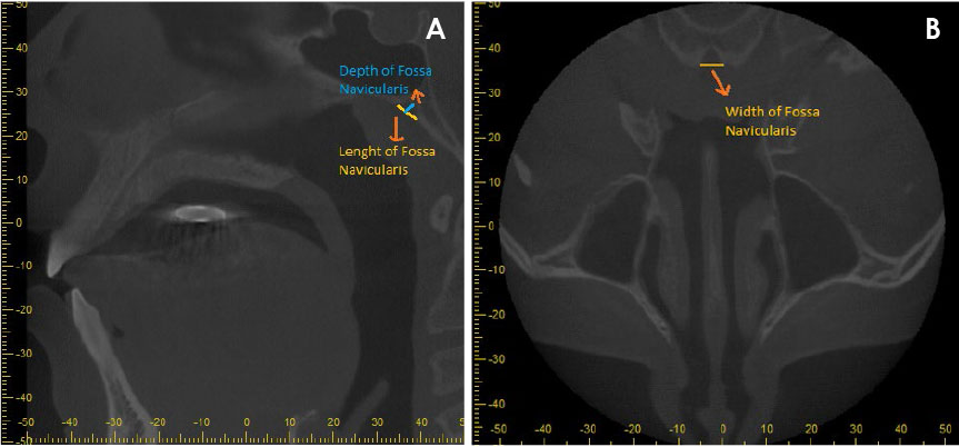
Fig. 1 A. Measurements of fossa navicularis depth and length on a sagittal image. B. Measurements of fossa navicularis width on an axial image.
Cited by 1 articles
-
Investigation of the prevalence and main features of skull-base anomalies and characteristics of the sphenoid sinus using cone-beam computed tomography
Aslıhan Akbulut, Oğuzhan Demirel, Kaan Orhan
J Korean Assoc Oral Maxillofac Surg. 2022;48(4):207-218. doi: 10.5125/jkaoms.2022.48.4.207.
Reference
-
1. Bayrak S, Göller Bulut D, Orhan K. Prevalence of anatomical variants in the clivus: fossa navicularis magna, canalis basilaris medianus, and craniopharyngeal canal. Surg Radiol Anat. 2019; 41:477–483.
Article2. Syed AZ, Mupparapu M. Fossa navicularis magna detection on cone-beam computed tomography. Imaging Sci Dent. 2016; 46:47–51.
Article3. Ersan N. Prevalence and morphometric features of fossa navicularis on cone beam computed tomography in Turkish population. Folia Morphol (Warsz). (in press).
Article4. Ahmad M, Jenny J, Downie M. Application of cone beam computed tomography in oral and maxillofacial surgery. Aust Dent J. 2012; 57 Suppl 1:82–94.
Article5. Ersan N. Prevalence of fossa navicularis among cleft palate patients detected by cone beam computed tomography. Yeditepe J Dent. 2017; 13:21–23.
Article6. Kizilkilic O, Yalcin O, Yildirim T, Sener L, Parmaksiz G, Erdogan B. Hypothalamic hamartoma associated with a craniopharyngeal canal. AJNR Am J Neuroradiol. 2005; 26:65–67.7. Pinilla-Arias D, Hinojosa J, Esparza J, Muñoz A. Recurrent meningitis and persistence of craniopharyngeal canal: case report. Neurocirugia (Astur). 2009; 20:50–53.
Article8. Abele TA, Salzman KL, Harnsberger HR, Glastonbury CM. Craniopharyngeal canal and its spectrum of pathology. AJNR Am J Neuroradiol. 2014; 35:772–777.
Article9. Currarino G. Canalis basilaris medianus and related defects of the basiocciput. AJNR Am J Neuroradiol. 1988; 9:208–211.10. Prabhu SP, Zinkus T, Cheng AG, Rahbar R. Clival osteomyelitis resulting from spread of infection through the fossa navicularis magna in a child. Pediatr Radiol. 2009; 39:995–998.
Article11. Segal N, Atamne E, Shelef I, Zamir S, Landau D. Intracranial infection caused by spreading through the fossa naviclaris magna - a case report and review of the literature. Int J Pediatr Otorhinolaryngol. 2013; 77:1919–1921.
Article12. Ray B, Kalthur SG, Kumar B, Bhat MR, D'souza AS, Gulati HS, et al. Morphological variations in the basioccipital region of the South Indian skull. Nepal J Med Sci. 2014; 3:124–128.
Article13. Cankal F, Ugur HC, Tekdemir I, Elhan A, Karahan T, Sevim A. Fossa navicularis: anatomic variation at the skull base. Clin Anat. 2004; 17:118–122.
Article14. Alalade AF, Briganti G, McKenzie JL, Gandhi M, Amato D, Panizza BJ, et al. Fossa navicularis in a pediatric patient: anatomical skull base variant with clinical implications. J Neurosurg Pediatr. 2018; 22:523–527.
Article15. Beltramello A, Puppini G, El-Dalati G, Girelli M, Cerini R, Sbarbati A, et al. Fossa navicularis magna. AJNR Am J Neuroradiol. 1998; 19:1796–1798.16. Akyel NG, Alımlı AG, Demirkan TH, Sivri M. Persistent craniopharyngeal canal, bilateral microphthalmia with colobomatous cysts, ectopic adenohypophysis with Rathke cleft cyst, and ectopic neurohypophysis: case report and review of the literature. Childs Nerv Syst. 2018; 34:1407–1410.
Article
- Full Text Links
-
- Actions
-
Cited
- CITED
-
- Close
- Share
-
- Similar articles
-
- Fossa navicularis magna detection on cone-beam computed tomography
- Mandibular condyle position in cone beam computed tomography
- Anomalies of the clivus of interest in dental practice: A systematic review
- Evaluation of the relation between the pulp stones and direct restorations using cone beam computed tomography in a Turkish subpopulation
- Three-dimensional imaging modalities in endodontics
