A Multicenter Prospective Validation Study for the Korean Thyroid Imaging Reporting and Data System in Patients with Thyroid Nodules
- Affiliations
-
- 1Department of Radiology, Ajou University School of Medicine, Suwon 16499, Korea.
- 2Department of Radiology, Konkuk University Medical Center, Konkuk University School of Medicine, Seoul 05030, Korea. mdmoonwj@naver.com
- 3Department of Radiology, Human Medical Imaging and Intervention Center, Seoul 06524, Korea.
- 4Department of Radiology, Ansan Hospital, Korea University School of Medicine, Ansan 15355, Korea.
- 5Department of Radiology, New Korea Hospital, Gimpo 10086, Korea.
- 6Department of Radiology, Chung-Ang University Hospital, Seoul 06973, Korea.
- KMID: 2458075
- DOI: http://doi.org/10.3348/kjr.2016.17.5.811
Abstract
OBJECTIVE
To validate a new risk stratification system for thyroid nodules, the Korean Thyroid Imaging Reporting and Data System (K-TIRADS), using a prospective design.
MATERIALS AND METHODS
From June 2013 to May 2015, 902 thyroid nodules were enrolled from four institutions. The type and predictive value of ultrasonography (US) predictors were analyzed according to the combination of the solidity and echogenicity of nodules; in addition, we determined malignancy risk and diagnostic performance for each category of K-TIRADS, and compared the efficacy of fine-needle aspiration (FNA) with a three-tier risk categorization system published in 2011.
RESULTS
The malignancy risk was significantly higher in solid hypoechoic nodules, as compared to partially cystic or isohyperechoic nodules (each p < 0.001). The presence of any suspicious US features had a significantly higher malignancy risk (73.4%) in solid hypoechoic nodules than in partially cystic or isohyperechoic nodules (4.3-38.5%; p < 0.001). The calculated malignancy risk in K-TIRADS categories 5, 4, 3, and 2 nodules were 73.4, 19.0, 3.5, and 0.0%, respectively; and the sensitivity, specificity, positive predictive value, negative predictive value, and accuracy for malignancy were 95.5, 58.6, 44.5, 96.9, and 69.5%, respectively, in K-TIRADS categories 4 and 5. The efficacy of FNA for detecting malignancy based on K-TIRADS was increased from 18.6% (101/544) to 22.5% (101/449), as compared with the three-tier risk categorization system (p < 0.001).
CONCLUSION
The proposed new risk stratification system based on solidity and echogenicity was useful for risk stratification of thyroid nodules and the decision for FNA. The malignancy risk of K-TIRADS was in agreement with the findings of a previous retrospective study.
Keyword
MeSH Terms
-
Adult
Biopsy, Fine-Needle/methods
Diagnosis, Differential
Female
Humans
Male
Middle Aged
Predictive Value of Tests
Prospective Studies
Research Design
Retrospective Studies
Risk Assessment/methods
Sensitivity and Specificity
Thyroid Neoplasms/diagnostic imaging/pathology
Thyroid Nodule/*diagnostic imaging/pathology
Ultrasonography/methods
Figure
-
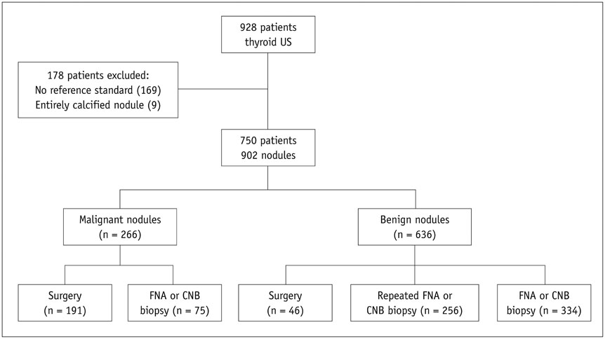
Fig. 1 Flow chart of study group. CNB = core needle biopsy, FNA = fine needle aspiration, US = ultrasonography
Cited by 7 articles
-
Concordance of Three International Guidelines for Thyroid Nodules Classified by Ultrasonography and Diagnostic Performance of Biopsy Criteria
Younghee Yim, Dong Gyu Na, Eun Ju Ha, Jung Hwan Baek, Jin Yong Sung, Ji-hoon Kim, Won-Jin Moon
Korean J Radiol. 2020;21(1):108-116. doi: 10.3348/kjr.2019.0215.Ultrasonographic Interval Changes in Solid Thyroid Nodules after Ultrasonography-Guided Fine-Needle Aspiration
Ik Jung Hwang, Dong Wook Kim, Yoo Jin Lee, Hye Jung Choo, Soo Jin Jung, Hye Jin Baek
Korean J Radiol. 2018;19(1):158-166. doi: 10.3348/kjr.2018.19.1.158.Ultrasonographic Echogenicity and Histopathologic Correlation of Thyroid Nodules in Core Needle Biopsy Specimens
Ji-hoon Kim, Dong Gyu Na, Hunkyung Lee
Korean J Radiol. 2018;19(4):673-681. doi: 10.3348/kjr.2018.19.4.673.Computer-Aided Diagnosis of Thyroid Nodules via Ultrasonography: Initial Clinical Experience
Young Jin Yoo, Eun Ju Ha, Yoon Joo Cho, Hye Lin Kim, Miran Han, So Young Kang
Korean J Radiol. 2018;19(4):665-672. doi: 10.3348/kjr.2018.19.4.665.Diagnostic Performance of a Combination of Shear Wave Elastography and B-Mode Ultrasonography in Differentiating Benign From Malignant Thyroid Nodules
Eung Koo Yeon, Yu-Mee Sohn, Mirinae Seo, Eui-Jong Kim, Young-Gyu Eun, Won Seo Park, Seong Jong Yun
Clin Exp Otorhinolaryngol. 2020;13(2):186-193. doi: 10.21053/ceo.2019.01235.Clinicopathological Characteristics and Recurrence-Free Survival of Rare Variants of Papillary Thyroid Carcinomas in Korea: A Retrospective Study
Mijin Kim, Sun Wook Cho, Young Joo Park, Hwa Young Ahn, Hee Sung Kim, Yong Joon Suh, Dughyun Choi, Bu Kyung Kim, Go Eun Yang, Il-Seok Park, Ka Hee Yi, Chan Kwon Jung, Bo Hyun Kim
Endocrinol Metab. 2021;36(3):619-627. doi: 10.3803/EnM.2021.974.Ultrasound Risk Stratification System of Thyroid Nodule
Hyun Jo Youn, Hyeong Eun Jeong, Ha Rim Ahn, Sang Yull Kang, Sung Hoo Jung
J Surg Ultrasound. 2023;10(2):25-31. doi: 10.46268/jsu.2023.10.2.25.
Reference
-
1. Haugen BR, Alexander EK, Bible KC, Doherty GM, Mandel SJ, Nikiforov YE, et al. 2015 American Thyroid Association Management Guidelines for Adult Patients with Thyroid Nodules and Differentiated Thyroid Cancer: The American Thyroid Association Guidelines Task Force on Thyroid Nodules and Differentiated Thyroid Cancer. Thyroid. 2016; 26:1–133.2. Moon WJ, Jung SL, Lee JH, Na DG, Baek JH, Lee YH, et al. Benign and malignant thyroid nodules: US differentiation--multicenter retrospective study. Radiology. 2008; 247:762–770.3. Gharib H, Papini E, Paschke R, Duick DS, Valcavi R, Hegedüs L, et al. American Association of Clinical Endocrinologists, Associazione Medici Endocrinologi, and European Thyroid Association Medical guidelines for clinical practice for the diagnosis and management of thyroid nodules: executive summary of recommendations. Endocr Pract. 2010; 16:468–475.4. Perros P, Boelaert K, Colley S, Evans C, Evans RM, Gerrard Ba G, et al. Guidelines for the management of thyroid cancer. Clin Endocrinol (Oxf). 2014; 81:Suppl 1. 1–122.5. Moon WJ, Baek JH, Jung SL, Kim DW, Kim EK, Kim JY, et al. Ultrasonography and the ultrasound-based management of thyroid nodules: consensus statement and recommendations. Korean J Radiol. 2011; 12:1–14.6. Wémeau JL, Sadoul JL, d'Herbomez M, Monpeyssen H, Tramalloni J, Leteurtre E, et al. Guidelines of the French society of endocrinology for the management of thyroid nodules. Ann Endocrinol (Paris). 2011; 72:251–228.7. Horvath E, Majlis S, Rossi R, Franco C, Niedmann JP, Castro A, et al. An ultrasonogram reporting system for thyroid nodules stratifying cancer risk for clinical management. J Clin Endocrinol Metab. 2009; 94:1748–1751.8. Park JY, Lee HJ, Jang HW, Kim HK, Yi JH, Lee W, et al. A proposal for a thyroid imaging reporting and data system for ultrasound features of thyroid carcinoma. Thyroid. 2009; 19:1257–1264.9. Kwak JY, Han KH, Yoon JH, Moon HJ, Son EJ, Park SH, et al. Thyroid imaging reporting and data system for US features of nodules: a step in establishing better stratification of cancer risk. Radiology. 2011; 260:892–899.10. Russ G, Royer B, Bigorgne C, Rouxel A, Bienvenu-Perrard M, Leenhardt L. Prospective evaluation of thyroid imaging reporting and data system on 4550 nodules with and without elastography. Eur J Endocrinol. 2013; 168:649–655.11. Cheng SP, Lee JJ, Lin JL, Chuang SM, Chien MN, Liu CL. Characterization of thyroid nodules using the proposed thyroid imaging reporting and data system (TI-RADS). Head Neck. 2013; 35:541–547.12. Kwak JY, Jung I, Baek JH, Baek SM, Choi N, Choi YJ, et al. Image reporting and characterization system for ultrasound features of thyroid nodules: multicentric Korean retrospective study. Korean J Radiol. 2013; 14:110–117.13. Russ G. Risk stratification of thyroid nodules on ultrasonography with the French TI-RADS: description and reflections. Ultrasonography. 2016; 35:25–38.14. Seo H, Na DG, Kim JH, Kim KW, Yoon JW. Ultrasound-based risk stratification for malignancy in thyroid nodules: a fourtier categorization system. Eur Radiol. 2015; 25:2153–2162.15. Na DG, Baek JH, Sung JY, Kim JH, Kim JK, Choi YJ, et al. Thyroid imaging reporting and data system risk stratification of thyroid nodules: categorization based on solidity and echogenicity. Thyroid. 2016; 26:562–572.16. Shin JH, Baek JH, Chung J, Ha EJ, Kim JH, Lee YH, et al. Ultrasonography diagnosis and imaging-based management of thyroid nodules: revised Korean society of thyroid radiology consensus statement and recommendations. Korean J Radiol. 2016; 17:370–395.17. Cibas ES, Ali SZ. The Bethesda system for reporting thyroid cytopathology. Thyroid. 2009; 19:1159–1165.18. Choi YJ, Baek JH, Hong MJ, Lee JH. Inter-observer variation in ultrasound measurement of the volume and diameter of thyroid nodules. Korean J Radiol. 2015; 16:560–565.19. Jung CK, Min HS, Park HJ, Song DE, Kim JH, Park SY, et al. Pathology reporting of thyroid core needle biopsy: a proposal of the Korean endocrine pathology thyroid core needle biopsy study group. J Pathol Transl Med. 2015; 49:288–299.20. Lee YH, Baek JH, Jung SL, Kwak JY, Kim JH, Shin JH, et al. Ultrasound-guided fine needle aspiration of thyroid nodules: a consensus statement by the korean society of thyroid radiology. Korean J Radiol. 2015; 16:391–401.21. Brito JP, Gionfriddo MR, Al Nofal A, Boehmer KR, Leppin AL, Reading C, et al. The accuracy of thyroid nodule ultrasound to predict thyroid cancer: systematic review and meta-analysis. J Clin Endocrinol Metab. 2014; 99:1253–1263.22. Na DG, Kim DS, Kim SJ, Ryoo JW, Jung SL. Thyroid nodules with isolated macrocalcification: malignancy risk and diagnostic efficacy of fine-needle aspiration and core needle biopsy. Ultrasonography. 2016; 35:212–219.23. Na DG, Kim JH, Kim DS, Kim SJ. Thyroid nodules with minimal cystic changes have a low risk of malignancy. Ultrasonography. 2016; 35:153–158.24. Kim DW, Lee EJ, In HS, Kim SJ. Sonographic differentiation of partially cystic thyroid nodules: a prospective study. AJNR Am J Neuroradiol. 2010; 31:1961–1966.25. Lee MJ, Kim EK, Kwak JY, Kim MJ. Partially cystic thyroid nodules on ultrasound: probability of malignancy and sonographic differentiation. Thyroid. 2009; 19:341–346.26. Park JM, Choi Y, Kwag HJ. Partially cystic thyroid nodules: ultrasound findings of malignancy. Korean J Radiol. 2012; 13:530–535.27. Seo HS, Lee DH, Park SH, Min HS, Na DG. Thyroid follicular neoplasms: can sonography distinguish between adenomas and carcinomas? J Clin Ultrasound. 2009; 37:493–500.28. Ozdemir D, Ersoy R, Cuhaci N, Arpaci D, Ersoy EP, Korukluoglu B, et al. Classical and follicular variant papillary thyroid carcinoma: comparison of clinical, ultrasonographical, cytological, and histopathological features in 444 patients. Endocr Pathol. 2011; 22:58–65.29. Rhee SJ, Hahn SY, Ko ES, Ryu JW, Ko EY, Shin JH. Follicular variant of papillary thyroid carcinoma: distinct biologic behavior based on ultrasonographic features. Thyroid. 2014; 24:683–688.
- Full Text Links
-
- Actions
-
Cited
- CITED
-
- Close
- Share
-
- Similar articles
-
- Thyroid Imaging Reporting and Data System (TIRADS)
- Korean Thyroid Imaging Reporting and Data System: Current Status, Challenges, and Future Perspectives
- 2021 Korean Thyroid Imaging Reporting and Data System (2021-K-TIRADS) and Imaging-Based Management of Thyroid Nodules: Korean Society of Thyroid Radiology Consensus Statement and Recommendations
- Clinical Application of the 2021 Korean Thyroid Imaging Reporting and Data System (K-TIRADS)
- Comparison of Korean vs. American Thyroid Imaging Reporting and Data System in Malignancy Risk Assessment of Indeterminate Thyroid Nodules
