Prospective Comparison of Liver Stiffness Measurements between Two Point Shear Wave Elastography Methods: Virtual Touch Quantification and Elastography Point Quantification
- Affiliations
-
- 1Department of Radiology, Seoul National University Hospital, Seoul 03080, Korea. jmsh@snu.ac.kr
- 2Institute of Radiation Medicine, Seoul National University Hospital, Seoul 03080, Korea.
- KMID: 2458067
- DOI: http://doi.org/10.3348/kjr.2016.17.5.750
Abstract
OBJECTIVE
To prospectively compare technical success rate and reliable measurements of virtual touch quantification (VTQ) elastography and elastography point quantification (ElastPQ), and to correlate liver stiffness (LS) measurements obtained by the two elastography techniques.
MATERIALS AND METHODS
Our study included 85 patients, 80 of whom were previously diagnosed with chronic liver disease. The technical success rate and reliable measurements of the two kinds of point shear wave elastography (pSWE) techniques were compared by χ2 analysis. LS values measured using the two techniques were compared and correlated via Wilcoxon signed-rank test, Spearman correlation coefficient, and 95% Bland-Altman limit of agreement. The intraobserver reproducibility of ElastPQ was determined by 95% Bland-Altman limit of agreement and intraclass correlation coefficient (ICC).
RESULTS
The two pSWE techniques showed similar technical success rate (98.8% for VTQ vs. 95.3% for ElastPQ, p = 0.823) and reliable LS measurements (95.3% for VTQ vs. 90.6% for ElastPQ, p = 0.509). The mean LS measurements obtained by VTQ (1.71 ± 0.47 m/s) and ElastPQ (1.66 ± 0.41 m/s) were not significantly different (p = 0.209). The LS measurements obtained by the two techniques showed strong correlation (r = 0.820); in addition, the 95% limit of agreement of the two methods was 27.5% of the mean. Finally, the ICC of repeat ElastPQ measurements was 0.991.
CONCLUSION
Virtual touch quantification and ElastPQ showed similar technical success rate and reliable measurements, with strongly correlated LS measurements. However, the two methods are not interchangeable due to the large limit of agreement.
MeSH Terms
Figure
-
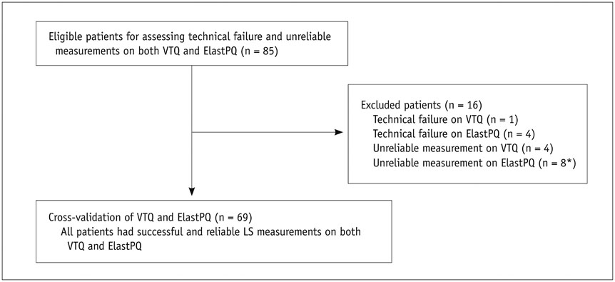
Fig. 1 Flow diagram of study population. *One patient who had technical failure on VTQ also had unreliable measurement on ElastPQ. ElastPQ = elastography point quantification, LS = liver stiffness, VTQ = virtual touch quantification
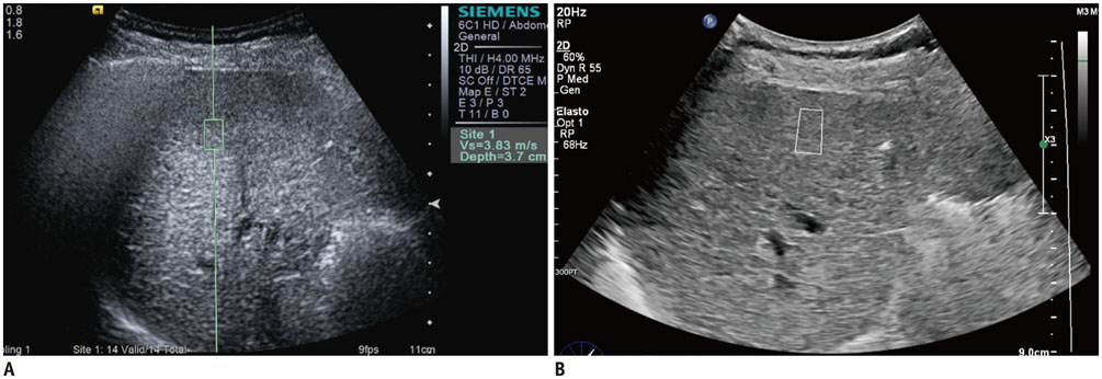
Fig. 2 LS measurements by two pSWE techniques. VTQ (A) and ElastPQ (B). For both measurements, operator placed predefined measuring box in liver. ElastPQ = elastography point quantification, LS = liver stiffness, pSWE = point shear wave elastography, VTQ = virtual touch quantification
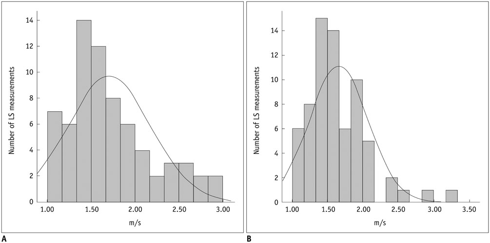
Fig. 3 Histogram showing distribution of mean LS measurements by two pSWE techniques. VTQ (A) and ElastPQ (B). Skewness/standard error of skewness was 3.10 for VTQ vs. 4.96 for ElastPQ. Black line = normal distribution fit. ElastPQ = elastography point quantification, LS = liver stiffness, pSWE = point shear wave elastography, VTQ = virtual touch quantification
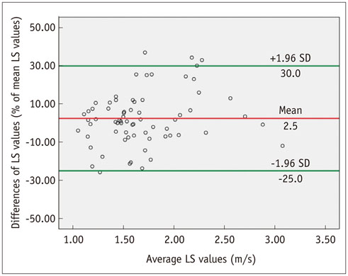
Fig. 4 Bland-Altman plot shows agreement in VTQ and ElastPQ LS measurements. X-axis shows means of repeated LS measurements, and y-axis shows difference between VTQ and ElastPQ LS measurements. Red line = mean difference, Green line = 95% limits of agreement. ElastPQ = elastography point quantification, LS = liver stiffness, VTQ = virtual touch quantification
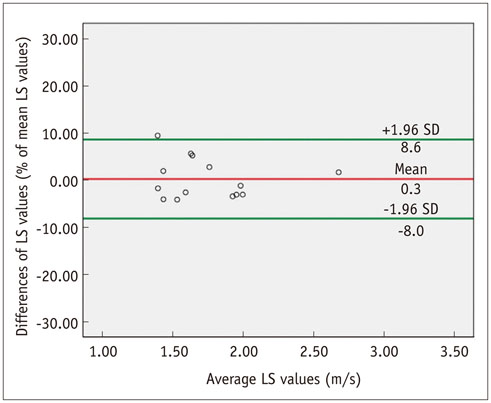
Fig. 5 Bland-Altman plot shows intraobserver agreement of ElastPQ measurement. X-axis shows means of repeated LS measurements, and y-axis shows difference between repeat ElastPQ LS measurements. Red line = mean difference, Green line = 95% limits of agreement. ElastPQ = elastography point quantification, LS = liver stiffness
Cited by 3 articles
-
Quantitative Measurement of Hepatic Fibrosis with Gadoxetic Acid-Enhanced Magnetic Resonance Imaging in Patients with Chronic Hepatitis B Infection: A Comparative Study on Aspartate Aminotransferase to Platelet Ratio Index and Fibrosis-4 Index
Guy Mok Lee, Youe Ree Kim, Jong Hyun Ryu, Tae-Hoon Kim, Eun Young Cho, Young Hwan Lee, Kwon-Ha Yoon
Korean J Radiol. 2017;18(3):444-451. doi: 10.3348/kjr.2017.18.3.444.Impact of Liver Fibrosis and Fatty Liver on T1rho Measurements: A Prospective Study
Shuangshuang Xie, Qing Li, Yue Cheng, Yu Zhang, Zhizheng Zhuo, Guiming Zhao, Wen Shen
Korean J Radiol. 2017;18(6):898-905. doi: 10.3348/kjr.2017.18.6.898.Validation of a New Point Shear-Wave Elastography Method for Noninvasive Assessment of Liver Fibrosis: A Prospective Multicenter Study
Ijin Joo, So Yeon Kim, Hee Sun Park, Eun Sun Lee, Hyo Jeong Kang, Jeong Min Lee
Korean J Radiol. 2019;20(11):1527-1535. doi: 10.3348/kjr.2019.0109.
Reference
-
1. Wong GL, Espinosa WZ, Wong VW. Personalized management of cirrhosis by non-invasive tests of liver fibrosis. Clin Mol Hepatol. 2015; 21:200–211.2. Rockey DC. Hepatic fibrosis, stellate cells, and portal hypertension. Clin Liver Dis. 2006; 10:459–479.3. Castera L. Invasive and non-invasive methods for the assessment of fibrosis and disease progression in chronic liver disease. Best Pract Res Clin Gastroenterol. 2011; 25:291–303.4. Bravo AA, Sheth SG, Chopra S. Liver biopsy. N Engl J Med. 2001; 344:495–500.5. Piccinino F, Sagnelli E, Pasquale G, Giusti G. Complications following percutaneous liver biopsy. A multicentre retrospective study on 68,276 biopsies. J Hepatol. 1986; 2:165–117.6. Wong GL. Prediction of fibrosis progression in chronic viral hepatitis. Clin Mol Hepatol. 2014; 20:228–236.7. Yoon JH, Lee JM, Joo I, Lee ES, Sohn JY, Jang SK, et al. Hepatic fibrosis: prospective comparison of MR elastography and US shear-wave elastography for evaluation. Radiology. 2014; 273:772–782.8. Jeong WK, Lim HK, Lee HK, Jo JM, Kim Y. Principles and clinical application of ultrasound elastography for diffuse liver disease. Ultrasonography. 2014; 33:149–160.9. Lee JE, Lee JM, Lee KB, Yoon JH, Shin CI, Han JK, et al. Noninvasive assessment of hepatic fibrosis in patients with chronic hepatitis B viral infection using magnetic resonance elastography. Korean J Radiol. 2014; 15:210–217.10. Kircheis G, Sagir A, Vogt C, Vom Dahl S, Kubitz R, Häussinger D. Evaluation of acoustic radiation force impulse imaging for determination of liver stiffness using transient elastography as a reference. World J Gastroenterol. 2012; 18:1077–1084.11. Shiina T, Nightingale KR, Palmeri ML, Hall TJ, Bamber JC, Barr RG, et al. WFUMB guidelines and recommendations for clinical use of ultrasound elastography: part 1: basic principles and terminology. Ultrasound Med Biol. 2015; 41:1126–1147.12. Tsochatzis EA, Gurusamy KS, Ntaoula S, Cholongitas E, Davidson BR, Burroughs AK. Elastography for the diagnosis of severity of fibrosis in chronic liver disease: a meta-analysis of diagnostic accuracy. J Hepatol. 2011; 54:650–659.13. Stebbing J, Farouk L, Panos G, Anderson M, Jiao LR, Mandalia S, et al. A meta-analysis of transient elastography for the detection of hepatic fibrosis. J Clin Gastroenterol. 2010; 44:214–219.14. Ma JJ, Ding H, Mao F, Sun HC, Xu C, Wang WP. Assessment of liver fibrosis with elastography point quantification technique in chronic hepatitis B virus patients: a comparison with liver pathological results. J Gastroenterol Hepatol. 2014; 29:814–819.15. Sandrin L, Fourquet B, Hasquenoph JM, Yon S, Fournier C, Mal F, et al. Transient elastography: a new noninvasive method for assessment of hepatic fibrosis. Ultrasound Med Biol. 2003; 29:1705–1713.16. Friedrich-Rust M, Nierhoff J, Lupsor M, Sporea I, Fierbinteanu-Braticevici C, Strobel D, et al. Performance of acoustic radiation force impulse imaging for the staging of liver fibrosis: a pooled meta-analysis. J Viral Hepat. 2012; 19:e212–e219.17. Bota S, Sporea I, Sirli R, Popescu A, Danila M, Costachescu D. Intra- and interoperator reproducibility of acoustic radiation force impulse (ARFI) elastography--preliminary results. Ultrasound Med Biol. 2012; 38:1103–1108.18. Ling W, Lu Q, Quan J, Ma L, Luo Y. Assessment of impact factors on shear wave based liver stiffness measurement. Eur J Radiol. 2013; 82:335–341.19. Chen S, Sanchez W, Callstrom MR, Gorman B, Lewis JT, Sanderson SO, et al. Assessment of liver viscoelasticity by using shear waves induced by ultrasound radiation force. Radiology. 2013; 266:964–970.20. Sporea I, Bota S, Grădinaru-Tas¸cău O, S¸irli R, Popescu A. Comparative study between two point shear wave elastographic techniques: acoustic radiation force impulse (ARFI) elastography and ElastPQ. Med Ultrason. 2014; 16:309–314.21. Musana KA, Yale SH, Abdulkarim AS. Tests of liver injury. Clin Med Res. 2004; 2:129–131.22. Ferraioli G, Filice C, Castera L, Choi BI, Sporea I, Wilson SR, et al. WFUMB guidelines and recommendations for clinical use of ultrasound elastography: part 3: liver. Ultrasound Med Biol. 2015; 41:1161–1179.23. Castéra L, Foucher J, Bernard PH, Carvalho F, Allaix D, Merrouche W, et al. Pitfalls of liver stiffness measurement: a 5-year prospective study of 13,369 examinations. Hepatology. 2010; 51:828–835.24. Zou KH, Tuncali K, Silverman SG. Correlation and simple linear regression. Radiology. 2003; 227:617–622.25. Bland JM, Altman DG. Statistical methods for assessing agreement between two methods of clinical measurement. Lancet. 1986; 1:307–310.26. Kim SY, Lee SS, Byun JH, Park SH, Kim JK, Park B, et al. Malignant hepatic tumors: short-term reproducibility of apparent diffusion coefficients with breath-hold and respiratory-triggered diffusion-weighted MR imaging. Radiology. 2010; 255:815–823.27. Boursier J, Zarski JP, de Ledinghen V, Rousselet MC, Sturm N, Lebail B, et al. Determination of reliability criteria for liver stiffness evaluation by transient elastography. Hepatology. 2013; 57:1182–1191.28. Chon YE, Choi EH, Song KJ, Park JY, Kim do Y, Han KH, et al. Performance of transient elastography for the staging of liver fibrosis in patients with chronic hepatitis B: a meta-analysis. PLoS One. 2012; 7:e44930.29. Ferraioli G, Tinelli C, Dal Bello B, Zicchetti M, Filice G, Filice C. Liver Fibrosis Study Group. Accuracy of real-time shear wave elastography for assessing liver fibrosis in chronic hepatitis C: a pilot study. Hepatology. 2012; 56:2125–2133.30. Guo Y, Parthasarathy S, Goyal P, McCarthy RJ, Larson AC, Miller FH. Magnetic resonance elastography and acoustic radiation force impulse for staging hepatic fibrosis: a meta-analysis. Abdom Imaging. 2015; 40:818–834.31. Nierhoff J, Chávez Ortiz AA, Herrmann E, Zeuzem S, Friedrich-Rust M. The efficiency of acoustic radiation force impulse imaging for the staging of liver fibrosis: a meta-analysis. Eur Radiol. 2013; 23:3040–3053.32. Ferraioli G, Tinelli C, Lissandrin R, Zicchetti M, Dal Bello B, Filice G, et al. Point shear wave elastography method for assessing liver stiffness. World J Gastroenterol. 2014; 20:4787–4796.
- Full Text Links
-
- Actions
-
Cited
- CITED
-
- Close
- Share
-
- Similar articles
-
- Inter-platform reproducibility of liver stiffness measured with two different point shear wave elastography techniques and 2-dimensional shear wave elastography using the comb-push technique
- Ultrasound Elastography for Liver Disease with Focus on Hepatic Fibrosis
- Diagnostic Performance of Quantitative Shear Wave Ultrasound Elastography for Thyroid Cancer
- Ultrasound-based Liver Elastography: Recent Advances
- Non-Invasive Liver Fibrosis Test Using Shear Wave Elastography
