Diffuse Infiltrative Splenic Lymphoma: Diagnostic Efficacy of Arterial-Phase CT
- Affiliations
-
- 1Department of Radiology, Chungnam National University Hospital, Chungnam National University College of Medicine, Daejeon 35015, Korea. jscho@cnu.ac.kr
- KMID: 2458065
- DOI: http://doi.org/10.3348/kjr.2016.17.5.734
Abstract
OBJECTIVE
To evaluate the diagnostic performance of obliteration of normal heterogeneous enhancement of the spleen (ONHES) on arterial phase (AP) computed tomography (CT) images in diffuse infiltrative splenic lymphoma (DISL).
MATERIALS AND METHODS
One hundred and thirty-six patients with lymphoma who had undergone two-phase (arterial and portal venous) abdominal CT were included in this study. We retrospectively evaluated the diagnostic performance of ONHES on AP CT in diagnosing DISL. Two observers evaluated ONHES on AP CT using the 5-point confidence level and assessed the presence or absence of subjective splenomegaly on axial CT images. Another two observers measured the splenic index as proposed by objective CT criteria. Statistical analysis included interobserver agreement and diagnostic performance of CT findings.
RESULTS
Eleven of the 136 patients with lymphoma had DISL. The area under the receiver operating characteristic curve of ONHES (0.948 for observer 1 and 0.922 for observer 2) was superior to that of the splenic index (0.872 for observer 3 and 0.877 for observer 4), but the difference was not statistically significant (p > 0.05). The diagnostic performance of ONHES in conjunction with subjective splenomegaly showed higher diagnostic performance, as compared with subjective splenomegaly alone (accuracy: 100% and 85.3% for observer 1, 98.5% and 87.5% for observer 2; positive predictive value: 100% and 35.5% for observer 1, 90.9% and 39.3% for observer 2, respectively).
CONCLUSION
Obliteration of normal heterogeneous enhancement of the spleen in conjunction with subjective splenomegaly can improve the diagnostic performance for DISL. Our results suggest that ONHES on AP CT images could be useful as an adjunctive diagnostic indicator of DISL in patients with lymphoma.
Keyword
MeSH Terms
-
Adult
Aged
Aged, 80 and over
Female
Humans
Image Interpretation, Computer-Assisted/methods
Lymphoma/complications/*diagnostic imaging
Male
Middle Aged
Observer Variation
Positron Emission Tomography Computed Tomography/methods
ROC Curve
Retrospective Studies
Splenic Neoplasms/complications/*diagnostic imaging
Splenomegaly/diagnostic imaging/etiology
Tomography, X-Ray Computed/methods
Young Adult
Figure
-
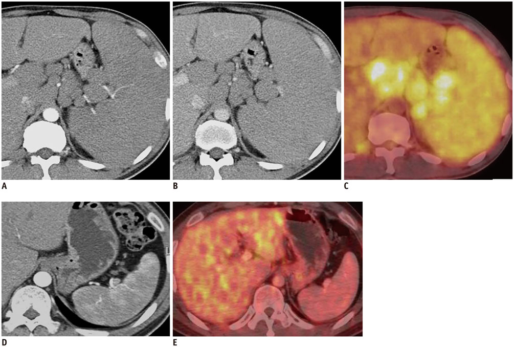
Fig. 1 41-year-old man with mantle cell lymphoma. Axial contrast-enhanced MDCT shows marked splenomegaly, multiple lymphadenopathies and obliteration of normal heterogeneous enhancement of spleen on AP image (A) and homogeneous enhancement on PVP image (B). PET/CT shows diffusely increased FDG uptake in spleen and multiple enlarged lymph nodes, suggesting lymphoma involvement (C). After chemotherapy, axial contrast-enhanced MDCT shows restoration of normal heterogeneous enhancement of spleen and interval marked decrease in size of enlarged spleen on AP image (D). Follow-up PET/CT after chemotherapy shows normal splenic uptake less than hepatic uptake (E). AP = arterial phase, FDG = fluorodeoxyglucose, MDCT = multidetector CT, PET/CT = positron emission tomography/CT, PVP = portal venous phase

Fig. 2 62-year-old man with diffuse large B-cell lymphoma. Axial contrast-enhanced MDCT shows obliteration of normal heterogeneous enhancement of spleen (ONHES) on AP image (A) and homogeneous enhancement on PVP image (B) with 247 cm3 of mean splenic index. However, there is no evidence of increased FDG uptake in spleen, suggesting false-positive finding for ONHES (C). AP = arterial phase, FDG = fluorodeoxyglucose, MDCT = multidetector CT, PVP = portal venous phase
Reference
-
1. Sandrasegaran K, Robinson PJ, Selby P. Staging of lymphoma in adults. Clin Radiol. 1994; 49:149–161.2. Saboo SS, Krajewski KM, O’Regan KN, Giardino A, Brown JR, Ramaiya N, et al. Spleen in haematological malignancies: spectrum of imaging findings. Br J Radiol. 2012; 85:81–92.3. Leite NP, Kased N, Hanna RF, Brown MA, Pereira JM, Cunha R, et al. Cross-sectional imaging of extranodal involvement in abdominopelvic lymphoproliferative malignancies. Radiographics. 2007; 27:1613–1634.4. Carroll BA, Ta HN. The ultrasonic appearance of extranodal abdominal lymphoma. Radiology. 1980; 136:419–425.5. Glazer GM, Axel L, Goldberg HI, Moss AA. Dynamic CT of the normal spleen. AJR Am J Roentgenol. 1981; 137:343–346.6. Donnelly LF, Foss JN, Frush DP, Bisset GS 3rd. Heterogeneous splenic enhancement patterns on spiral CT images in children: minimizing misinterpretation. Radiology. 1999; 210:493–497.7. Miles KA, McPherson SJ, Hayball MP. Transient splenic inhomogeneity with contrast-enhanced CT: mechanism and effect of liver disease. Radiology. 1995; 194:91–95.8. Prassopoulos P, Daskalogiannaki M, Raissaki M, Hatjidakis A, Gourtsoyiannis N. Determination of normal splenic volume on computed tomography in relation to age, gender and body habitus. Eur Radiol. 1997; 7:246–248.9. Frank K, Linhart P, Kortsik C, Wohlenberg H. [Sonographic determination of spleen size: normal dimensions in adults with a healthy spleen]. Ultraschall Med. 1986; 7:134–137.10. Lackner K, Brecht G, Janson R, Scherholz K, Lützeler A, Thurn P. [The value of computer tomography in the staging of primary lymph node neoplasms (author’s transl)]. Rofo. 1980; 132:21–30.11. Strijk SP, Wagener DJ, Bogman MJ, de Pauw BE, Wobbes T. The spleen in Hodgkin disease: diagnostic value of CT. Radiology. 1985; 154:753–757.12. Rini JN, Leonidas JC, Tomas MB, Palestro CJ. 18F-FDG PET versus CT for evaluating the spleen during initial staging of lymphoma. J Nucl Med. 2003; 44:1072–1074.13. Rini JN, Manalili EY, Hoffman MA, Karayalcin G, Mehrotra B, Tomas MB, et al. F-18 FDG versus Ga-67 for detecting splenic involvement in Hodgkin’s disease. Clin Nucl Med. 2002; 27:572–577.14. Hernandez-Maraver D, Hernandez-Navarro F, Gomez-Leon N, Coya J, Rodriguez-Vigil B, Madero R, et al. Positron emission tomography/computed tomography: diagnostic accuracy in lymphoma. Br J Haematol. 2006; 135:293–302.15. Hanley JA, McNeil BJ. A method of comparing the areas under receiver operating characteristic curves derived from the same cases. Radiology. 1983; 148:839–843.16. Gorg C, Weide R, Schwerk WB. Malignant splenic lymphoma: sonographic patterns, diagnosis and follow-up. Clin Radiol. 1997; 52:535–540.17. Goerg C, Schwerk WB, Goerg K, Havemann K. Sonographic patterns of the affected spleen in malignant lymphoma. J Clin Ultrasound. 1990; 18:569–574.18. Hess CF, Kurtz B, Hoffmann W, Bamberg M. Ultrasound diagnosis of splenic lymphoma: ROC analysis of multidimensional splenic indices. Br J Radiol. 1993; 66:859–864.19. de Jong PA, van Ufford HM, Baarslag HJ, de Haas MJ, Wittebol SH, Quekel LG, et al. CT and 18F-FDG PET for noninvasive detection of splenic involvement in patients with malignant lymphoma. AJR Am J Roentgenol. 2009; 192:745–753.20. Gartner LP, Hiatt JL. Color textbook of histology. 2nd ed. Philadelphia: Saunders;2001. p. 291–294.21. Bhatia K, Sahdev A, Reznek RH. Lymphoma of the spleen. Semin Ultrasound CT MR. 2007; 28:12–20.22. Guermazi A, Brice P, de Kerviler E E, Fermé C, Hennequin C, Meignin V, et al. Extranodal Hodgkin disease: spectrum of disease. Radiographics. 2001; 21:161–179.23. Görg C, Faoro C, Bert T, Tebbe J, Neesse A, Wilhelm C. Contrast enhanced ultrasound of splenic lymphoma involvement. Eur J Radiol. 2011; 80:169–174.24. Weissleder R, Elizondo G, Stark DD, Hahn PF, Marfil J, Gonzalez JF, et al. The diagnosis of splenic lymphoma by MR imaging: value of superparamagnetic iron oxide. AJR Am J Roentgenol. 1989; 152:175–180.25. Munker R, Stengel A, Stäbler A, Hiller E, Brehm G. Diagnostic accuracy of ultrasound and computed tomography in the staging of Hodgkin’s disease. Verification by laparotomy in 100 cases. Cancer. 1995; 76:1460–1146.26. Cheson BD, Pfistner B, Juweid ME, Gascoyne RD, Specht L, Horning SJ, et al. Revised response criteria for malignant lymphoma. J Clin Oncol. 2007; 25:579–586.27. Fishman EK, Kuhlman JE, Jones RJ. CT of lymphoma: spectrum of disease. Radiographics. 1991; 11:647–669.
- Full Text Links
-
- Actions
-
Cited
- CITED
-
- Close
- Share
-
- Similar articles
-
- Primary Splenic Lymphoma with Splenic Hilar Lymphadenopathy
- Transient Inhomogeneous Contrast Enhancement of the Spleen on Arterial Phase of Spiral CT
- Hypotensive Splenic Infarction: A Case Report
- CT Findings of Malarial Spleens
- Diffuse Infiltrative Primary Cardiac Lymphoma with Delayed Extracardiac Involvement
