Radiologic Diagnosis of Asbestosis in Korea
- Affiliations
-
- 1Department of Radiology, Dongguk University Ilsan Hospital, Dongguk University College of Medicine, Goyang 10326, Korea. jeungkim@dumc.or.kr
- 2Department of Radiology, Ewha Womans University Mokdong Hospital, Ewha Womans University School of Medicine, Seoul 07985, Korea.
- 3Department of Radiology, Gachon University Gil Medical Center, Gachon University, Incheon 21565, Korea.
- KMID: 2458059
- DOI: http://doi.org/10.3348/kjr.2016.17.5.674
Abstract
- Asbestosis is the most important change noted in the lung parenchyma after environmental and occupational exposure to asbestos fibers. It is characterized by diffuse interstitial pulmonary fibrosis. In Korea, the incidence of asbestosis will continue to increase for many years to come and the government enacted the Asbestos Damage Relief Law in 2011 to provide compensation to those suffering from asbestos-related diseases. Radiologic evaluation is necessary for diagnosis of asbestosis, and radiologists play a key role in this process. Therefore, it is important for radiologists to be aware of the various imaging features of asbestosis.
Keyword
MeSH Terms
Figure
-
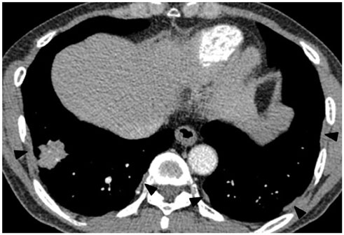
Fig. 1 56-year-old male resident for 15 years near asbestos mine with work history at construction company for 10 years. Chest CT shows irregular lobulated enhancing nodule in right lower lobe, which are confirmed as lung adenocarcinoma. There are multiple discrete pleural plaques (arrowheads) in both lower hemithoraces.
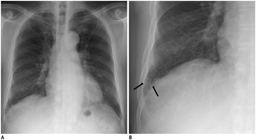
Fig. 2 74-year-old male resident for 33 years under asbestos roof. A. Chest PA radiograph shows reticular opacities in both lower lungs. B. Magnification of right lower lung shows reticular and small nodular opacities (arrows). PA = posteroanterior
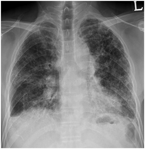
Fig. 3 57-year-old male resident near asbestos mine since birth. Chest PA radiograph shows reticular densities and honeycomb cysts in both lungs. PA = posteroanterior
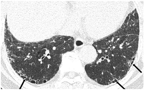
Fig. 4 CT findings of patient of Figure 2. Chest CT shows honeycomb cysts (arrows) in subpleural portions of both lower lobes.

Fig. 5 73-year-old female resident for 34 years near asbestos mine. CT shows obvious (arrows) and faint (arrowheads) dot-like opacities in subpleural portions of lower lung.
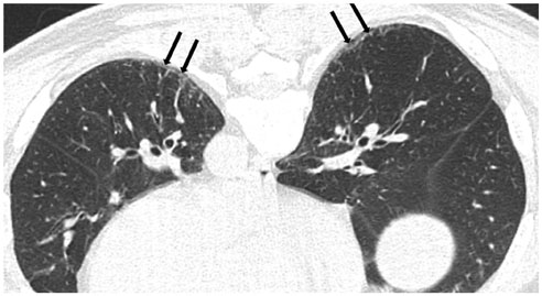
Fig. 6 60-year-old male resident near asbestos mine since birth with 3 year work history at mine. Prone CT reveals subpleural curvilinear lines (arrows) in lower lungs.
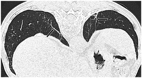
Fig. 7 82-year-old male resident near asbestos mine since birth. Prone CT reveals parenchymal bands (arrows) extending through lung to contact pleural surface.
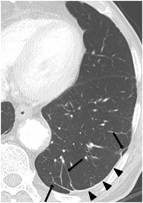
Fig. 8 79-year-old male resident for 43 years near asbestos mine. CT shows multiple parenchymal bands (arrows) adjacent to pleural plaques (arrowheads) in left lower lobe.
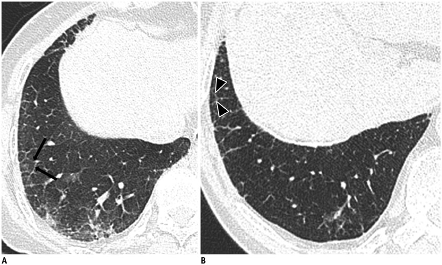
Fig. 9 79-year-old female resident for 60 years near asbestos mine with 5 year work history at mine. A, B. CT shows interlobular septal thickening (arrows) (A) and intralobular interstitial thickening (arrowheads) (B) connected to peripheral pulmonary arteries.
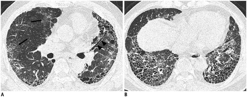
Fig. 10 69-year-old male with 20 year work history at construction company. A. CT shows ground-glass opacity (arrows), traction bronchiectasis (arrowheads) and honeycomb cysts. B. CT shows multilayered honeycomb cysts in both lower lungs.
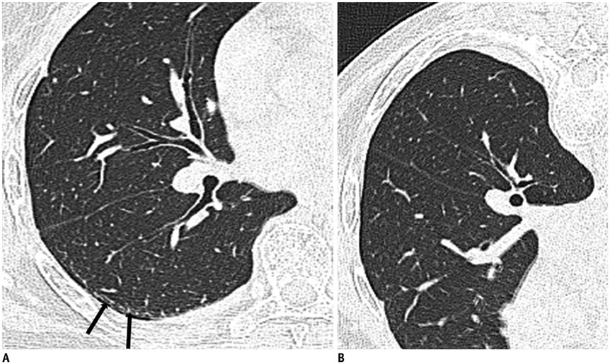
Fig. 11 Dependent atelectasis in patient with history of asbestos exposure. A. On supine CT, subpleural line is visible in posterior portion of superior segment of right lower lobe (arrows). B. On prone CT, subpleural line is disappeared, indicative that thin posterior opacity is dependent atelectasis.
Reference
-
1. Cugell DW, Kamp DW. Asbestos and the pleura: a review. Chest. 2004; 125:1103–1117.2. Kim HR. Overview of asbestos issues in Korea. J Korean Med Sci. 2009; 24:363–367.3. Roach HD, Davies GJ, Attanoos R, Crane M, Adams H, Phillips S. Asbestos: when the dust settles an imaging review of asbestos-related disease. Radiographics. 2002; 22(Spec No):S167–S184.4. Lynch DA, Gamsu G, Ray CS, Aberle DR. Asbestos-related focal lung masses: manifestations on conventional and high-resolution CT scans. Radiology. 1988; 169:603–607.5. Kim JS, Lynch DA. Imaging of nonmalignant occupational lung disease. J Thorac Imaging. 2002; 17:238–260.6. Gefter WB, Epstein DM, Miller WT. Radiographic evaluation of asbestos-related chest disorders. Crit Rev Diagn Imaging. 1984; 21:133–181.7. Gefter WB, Conant EF. Issues and controversies in the plain-film diagnosis of asbestos-related disorders in the chest. J Thorac Imaging. 1988; 3:11–28.8. Clements M, Berry G, Shi J, Ware S, Yates D, Johnson A. Projected mesothelioma incidence in men in New South Wales. Occup Environ Med. 2007; 64:747–752.9. Hodgson JT, McElvenny DM, Darnton AJ, Price MJ, Peto J. The expected burden of mesothelioma mortality in Great Britain from 2002 to 2050. Br J Cancer. 2005; 92:587–593.10. Paek DM. Asbestos problems of Korea. Environ Law Policy. 2009; 2:5–22.11. Churg A, Wright JL, DePaoli L, Wiggs B. Mineralogic correlates of fibrosis in chrysotile miners and millers. Am Rev Respir Dis. 1989; 139:891–896.12. Churg A, Wright J, Wiggs B, Depaoli L. Mineralogic parameters related to amosite asbestos-induced fibrosis in humans. Am Rev Respir Dis. 1990; 142(6 Pt 1):1331–1336.13. Wright JL, Cagle P, Churg A, Colby TV, Myers J. Diseases of the small airways. Am Rev Respir Dis. 1992; 146:240–262.14. Bégin R, Cantin A, Berthiaume Y, Boileau R, Péloquin S, Massé S. Airway function in lifetime-nonsmoking older asbestos workers. Am J Med. 1983; 75:631–638.15. Craighead JE, Abraham JL, Churg A, Green FH, Kleinerman J, Pratt PC, et al. The pathology of asbestos-associated diseases of the lungs and pleural cavities: diagnostic criteria and proposed grading schema. Report of the Pneumoconiosis Committee of the College of American Pathologists and the National Institute for Occupational Safety and Health. Arch Pathol Lab Med. 1982; 106:544–596.16. Green FH, Attfield M. Pathology standards for asbestosis. Scand J Work Environ Health. 1983; 9(2 Spec No):162–168.17. American Thoracic Society. Diagnosis and initial management of nonmalignant diseases related to asbestos. Am J Respir Crit Care Med. 2004; 170:691–715.18. Wolff H, Vehmas T, Oksa P, Rantanen J, Vainio H. Asbestos, asbestosis, and cancer, the Helsinki criteria for diagnosis and attribution 2014: recommendations. Scand J Work Environ Health. 2015; 41:5–15.19. Roggli VL, Gibbs AR, Attanoos R, Churg A, Popper H, Cagle P, et al. Pathology of asbestosis-an update of the diagnostic criteria: report of the asbestosis committee of the college of american pathologists and pulmonary pathology society. Arch Pathol Lab Med. 2010; 134:462–480.20. Suganuma N, Kusaka Y, Hering KG, Vehmas T, Kraus T, Arakawa H, et al. Reliability of the proposed international classification of high-resolution computed tomography for occupational and environmental respiratory diseases. J Occup Health. 2009; 51:210–222.21. Prazakova S, Thomas PS, Sandrini A, Yates DH. Asbestos and the lung in the 21st century: an update. Clin Respir J. 2014; 8:1–10.22. Weiss W. Asbestosis: a marker for the increased risk of lung cancer among workers exposed to asbestos. Chest. 1999; 115:536–549.23. Hillerdal G. Malignant mesothelioma 1982: review of 4710 published cases. Br J Dis Chest. 1983; 77:321–343.24. Kim HR, Ahn YS, Jung SH. Epidemiologic characteristics of malignant mesothelioma in Korea. J Korean Med Assoc. 2009; 52:449–455.25. Raffaelli I, Festa G, Costantini AS, Leva G, Gorini G. [Mortality in a cohort of asbestos cement workers in Carrara, Italy]. Med Lav. 2007; 98:156–163.26. Luberto F, Amendola P, Belli S, Bruno C, Candela S, Grignoli M, et al. [Mortality study of asbestos cement workers in Emilia-Romagna]. Epidemiol Prev. 2004; 28:239–246.27. Gallus S, Pacifici R, Colombo P, Scarpino V, Zuccaro P, Bosetti C, et al. Prevalence of smoking and attitude towards smoking regulation in Italy, 2004. Eur J Cancer Prev. 2006; 15:77–81.28. Bałczewska E. Smoking and tobacco control in Poland. Eur J Dent Educ. 2004; 8:Suppl 4. 42–45.29. Chen M, Tse LA, Au RK, Yu IT, Wang XR, Lao XQ, et al. Mesothelioma and lung cancer mortality: a historical cohort study among asbestosis workers in Hong Kong. Lung Cancer. 2012; 76:165–170.30. Chong S, Lee KS, Chung MJ, Han J, Kwon OJ, Kim TS. Pneumoconiosis: comparison of imaging and pathologic findings. Radiographics. 2006; 26:59–77.31. Kipen HM, Lilis R, Suzuki Y, Valciukas JA, Selikoff IJ. Pulmonary fibrosis in asbestos insulation workers with lung cancer: a radiological and histopathological evaluation. Br J Ind Med. 1987; 44:96–100.32. Zachrisson S, Vikgren J, Svalkvist A, Johnsson AA, Boijsen M, Flinck A, et al. Effect of clinical experience of chest tomosynthesis on detection of pulmonary nodules. Acta Radiol. 2009; 50:884–891.33. Vikgren J, Zachrisson S, Svalkvist A, Johnsson AA, Boijsen M, Flinck A, et al. Comparison of chest tomosynthesis and chest radiography for detection of pulmonary nodules: human observer study of clinical cases. Radiology. 2008; 249:1034–1041.34. Quaia E, Baratella E, Cioffi V, Bregant P, Cernic S, Cuttin R, et al. The value of digital tomosynthesis in the diagnosis of suspected pulmonary lesions on chest radiography: analysis of diagnostic accuracy and confidence. Acad Radiol. 2010; 17:1267–1274.35. Jung HN, Chung MJ, Koo JH, Kim HC, Lee KS. Digital tomosynthesis of the chest: utility for detection of lung metastasis in patients with colorectal cancer. Clin Radiol. 2012; 67:232–238.36. McLoud TC. Conventional radiography in the diagnosis of asbestos-related disease. Radiol Clin North Am. 1992; 30:1177–1189.37. Lee G, Jeong YJ, Kim KI, Song JW, Kang DM, Kim YD, et al. Comparison of chest digital tomosynthesis and chest radiography for detection of asbestos-related pleuropulmonary disease. Clin Radiol. 2013; 68:376–382.38. Staples CA, Gamsu G, Ray CS, Webb WR. High resolution computed tomography and lung function in asbestos-exposed workers with normal chest radiographs. Am Rev Respir Dis. 1989; 139:1502–1508.39. Friedman AC, Fiel SB, Fisher MS, Radecki PD, Lev-Toaff AS, Caroline DF. Asbestos-related pleural disease and asbestosis: a comparison of CT and chest radiography. AJR Am J Roentgenol. 1988; 150:269–275.40. Tekath M, Dutheil F, Bellini R, Roche A, Pereira B, Naughton G, et al. Comparison of the ultra-low-dose Veo algorithm with the gold standard filtered back projection for detecting pulmonary asbestos-related conditions: a clinical observational study. BMJ Open. 2014; 4:e004980.41. Akira M, Yokoyama K, Yamamoto S, Higashihara T, Morinaga K, Kita N, et al. Early asbestosis: evaluation with high-resolution CT. Radiology. 1991; 178:409–416.42. Akira M, Yamamoto S, Yokoyama K, Kita N, Morinaga K, Higashihara T, et al. Asbestosis: high-resolution CT-pathologic correlation. Radiology. 1990; 176:389–394.43. Friedman AC, Fiel SB, Radecki PD, Lev-Toaff AS. Computed tomography of benign pleural and pulmonary parenchymal abnormalities related to asbestos exposure. Semin Ultrasound CT MR. 1990; 11:393–408.44. Aberle DR, Gamsu G, Ray CS. High-resolution CT of benign asbestos-related diseases: clinical and radiographic correlation. AJR Am J Roentgenol. 1988; 151:883–891.45. Aberle DR, Gamsu G, Ray CS, Feuerstein IM. Asbestos-related pleural and parenchymal fibrosis: detection with high-resolution CT. Radiology. 1988; 166:729–734.46. Akira M, Yamamoto S, Inoue Y, Sakatani M. High-resolution CT of asbestosis and idiopathic pulmonary fibrosis. AJR Am J Roentgenol. 2003; 181:163–169.47. al-Jarad N, Strickland B, Pearson MC, Rubens MB, Rudd RM. High resolution computed tomographic assessment of asbestosis and cryptogenic fibrosing alveolitis: a comparative study. Thorax. 1992; 47:645–650.48. Gevenois PA, de Maertelaer V, Madani A, Winant C, Sergent G, De Vuyst P. Asbestosis, pleural plaques and diffuse pleural thickening: three distinct benign responses to asbestos exposure. Eur Respir J. 1998; 11:1021–1027.49. Staples CA. Computed tomography in the evaluation of benign asbestos-related disorders. Radiol Clin North Am. 1992; 30:1191–1207.50. Lee EK, Kim JS, Kim Y, Park JS. CT Findings in people who were environmentally exposed to asbestos in Korea. J Korean Med Sci. 2015; 30:1896–1901.51. Kim Y, Myong JP, Lee JK, Kim JS, Kim YK, Jung SH. CT Characteristics of pleural plaques related to occupational or environmental asbestos exposure from South Korean asbestos mines. Korean J Radiol. 2015; 16:1142–1152.52. Copley SJ, Wells AU, Sivakumaran P, Rubens MB, Lee YC, Desai SR, et al. Asbestosis and idiopathic pulmonary fibrosis: comparison of thin-section CT features. Radiology. 2003; 229:731–736.53. Arakawa H, Kishimoto T, Ashizawa K, Kato K, Okamoto K, Honma K, et al. Asbestosis and other pulmonary fibrosis in asbestos-exposed workers: high-resolution CT features with pathological correlations. Eur Radiol. 2016; 26:1485–1492.
