Self-Expandable Stents in Vascular Stenosis of Moderate to Large-Sized Vessels in Congenital Heart Disease: Early and Intermediate-Term Results
- Affiliations
-
- 1Department of Pediatrics, Korea University Hospital, Ansan, Korea. jgynhg@naver.com
- KMID: 2458000
- DOI: http://doi.org/10.4070/kcj.2019.0067
Abstract
- BACKGROUND AND OBJECTIVES
Vascular stenosis after surgical repair frequently occurs in congenital heart disease. Although conventional balloon dilation is a useful option for stenotic lesions, restenosis may occur. Consequently, balloon expandable stents have been used; however, there are a limited number of balloon expandable stents in our country. Here, we report the early and intermediate-term outcomes of self-expandable stents in vascular stenosis of moderate to large-sized vessels in congenital heart disease.
METHODS
Twelve self-expandable stents were implanted in 9 patients between February 2012 and January 2019. The median age and weight were 12 years (range, 4-39 years) and 38 kg (range, 19-69 kg), respectively. The patients were followed-up for a median duration of 43 months (range, 1-83 months) after stent implantation.
RESULTS
Nine self-expandable stents were implanted in the pulmonary artery, 2 stents in the right ventricle to the pulmonary artery conduit, and 1 stent in the coarctation. The narrowest diameter of the stented vessel increased from 5.7±3.2 mm to 12.6±3.4 mm (p<0.05). The mean pressure gradient across the stenotic lesion decreased from 23.0±28.2 mmHg to 3.2±3.6 mmHg (p<0.05). Distal migration of the stent occurred in 1 patient, and significant neointimal ingrowth was noted in 1 patient.
CONCLUSIONS
The self-expandable stent may be a useful option to relieve vascular stenosis in moderate to large-sized vessels with acceptable intermediate-term outcomes.
Keyword
MeSH Terms
Figure
-

Figure 1 (A) Catheterization after Fontan operation demonstrated multiple narrowings of the LPA. (B) Angiography after 2 self-expandable stent implantation showing relief of the stenosis of LPA. LPA = left pulmonary artery.
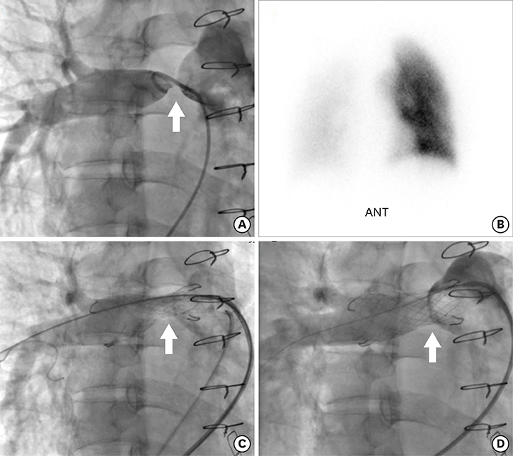
Figure 2 (A) Angiography showing severe stenosis of the right pulmonary artery (arrow). (B) Lung perfusion scan showing right-to-left lung perfusion ratio of 15/85. (C) Immediate follow-up angiography after stent implantation. (D) After 6 months of stent placement, there was near-complete relief of the stenosis by constant radial force. ANT = anterior.
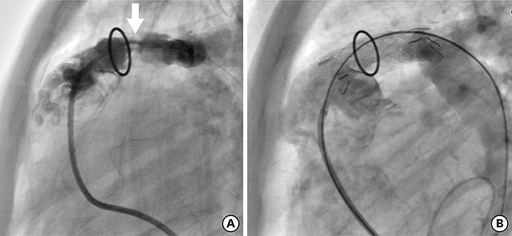
Figure 3 (A) Catheterization after Rastelli operation demonstrated severe stenosis of right ventricle to pulmonary artery conduit (arrow). (B) Angiography after self-expandable stent implantation with 14 mm in diameter showing near-complete relief of the stenosis in RV-PA conduit.

Figure 4 (A) Angiography after coarctoplasty showing focal stenosis in descending aorta. (B) A computed tomography after self-expandable stent implantation showing near-complete relief of the stenosis.
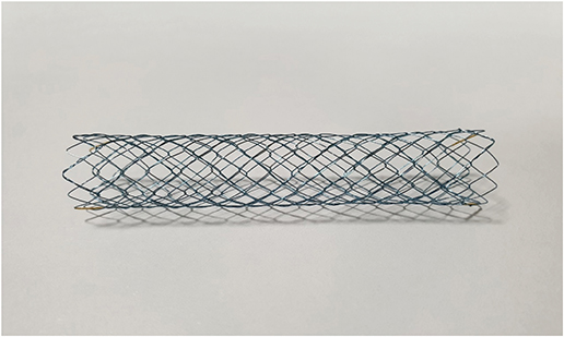
Figure 5 Hercules self-expandable vascular stent, bare metal stent which is made in a shape of memory nitinol. It has soft ends and good flexibility.
Cited by 2 articles
-
The Significance of Self-Expandable Stents in Patients with Congenital Heart Disease in Current Era
Sang-Yun Lee
Korean Circ J. 2019;49(10):943-944. doi: 10.4070/kcj.2019.0201.Endovascular Therapy of Iliac Artery Disease: Stent Matters
Su Hong Kim
Korean Circ J. 2021;51(5):452-454. doi: 10.4070/kcj.2021.0022.
Reference
-
1. Haji Zeinali AM, Sadeghian M, Qureshi SA, Ghazi P. Midterm to long-term safety and efficacy of self-expandable nitinol stent implantation for coarctation of aorta in adults. Catheter Cardiovasc Interv. 2017; 90:425–431.
Article2. Goreczny S, Qureshi SA, Rosenthal E, et al. Comparison of self-expandable and balloon-expanding stents for hybrid ductal stenting in hypoplastic left heart complex. Cardiol Young. 2017; 27:837–845.
Article3. Cheung YF, Sanatani S, Leung MP, Human DG, Chau AK, Culham JA. Early and intermediate-term complications of self-expanding stents limit its potential application in children with congenital heart disease. J Am Coll Cardiol. 2000; 35:1007–1015.
Article4. Redington AN, Weil J, Somerville J. Self expanding stents in congenital heart disease. Br Heart J. 1994; 72:378–383.
Article5. Pavithran S, Bhattacharjya S, Chandrasekaran R, Sivakumar K. Stent angioplasty of narrowed right ventricular outflow conduits and pulmonary arteries consistently reduces right ventricular systolic pressures and delays subsequent surgeries. Indian Heart J. 2018; 70:879–886.
Article6. Adjagba PM, Hanna B, Miró J, et al. Percutaneous angioplasty used to manage native and recurrent coarctation of the aorta in infants younger than 1 year: immediate and midterm results. Pediatr Cardiol. 2014; 35:1155–1161.7. Gonzalez I, Kenny D, Slyder S, Hijazi ZM. Medium and long-term outcomes after bilateral pulmonary artery stenting in children and adults with congenital heart disease. Pediatr Cardiol. 2013; 34:179–184.
Article8. Forbes TJ, Garekar S, Amin Z, et al. Procedural results and acute complications in stenting native and recurrent coarctation of the aorta in patients over 4 years of age: a multi-institutional study. Catheter Cardiovasc Interv. 2007; 70:276–285.
Article9. Mullins CE, O'Laughlin MP, Vick GW 3rd, et al. Implantation of balloon-expandable intravascular grafts by catheterization in pulmonary arteries and systemic veins. Circulation. 1988; 77:188–199.
Article10. O'Laughlin MP, Perry SB, Lock JE, Mullins CE. Use of endovascular stents in congenital heart disease. Circulation. 1991; 83:1923–1939.11. Gendera K, Ewert P, Tanase D, et al. Balloon-expandable stents for recoarctation of the aorta in small children. Two centre experience. Int J Cardiol. 2018; 263:34–39.
Article12. Quandt D, Ramchandani B, Bhole V, et al. Initial experience with the cook formula balloon expandable stent in congenital heart disease. Catheter Cardiovasc Interv. 2015; 85:259–266.
Article13. Chakrabarti S, Kenny D, Morgan G, et al. Balloon expandable stent implantation for native and recurrent coarctation of the aorta--prospective computed tomography assessment of stent integrity, aneurysm formation and stenosis relief. Heart. 2010; 96:1212–1216.
Article14. Krisnanda C, Menahem S, Lane GK. Intravascular stent implantation for the management of pulmonary artery stenosis. Heart Lung Circ. 2013; 22:56–70.
Article15. Okubo M, Benson LN. Intravascular and intracardiac stents used in congenital heart disease. Curr Opin Cardiol. 2001; 16:84–91.
Article16. Peters B, Ewert P, Berger F. The role of stents in the treatment of congenital heart disease: current status and future perspectives. Ann Pediatr Cardiol. 2009; 2:3–23.
Article17. O'Laughlin MP, Slack MC, Grifka RG, Perry SB, Lock JE, Mullins CE. Implantation and intermediate-term follow-up of stents in congenital heart disease. Circulation. 1993; 88:605–614.18. van Gameren M, Witsenburg M, Takkenberg JJ, et al. Early complications of stenting in patients with congenital heart disease: a multicentre study. Eur Heart J. 2006; 27:2709–2715.
Article19. Powell AJ, Lock JE, Keane JF, Perry SB. Prolongation of RV-PA conduit life span by percutaneous stent implantation. Intermediate-term results. Circulation. 1995; 92:3282–3288.20. Frias PA, Meranze SG, Graham TP Jr, Doyle TP. Relief of right ventricular to pulmonary artery conduit stenosis using a self-expanding stent. Catheter Cardiovasc Interv. 1999; 47:52–54.
Article21. Cowley CG, Orsmond GS, Feola P, McQuillan L, Shaddy RE. Long-term, randomized comparison of balloon angioplasty and surgery for native coarctation of the aorta in childhood. Circulation. 2005; 111:3453–3456.
Article22. Rodés-Cabau J, Miró J, Dancea A, et al. Comparison of surgical and transcatheter treatment for native coarctation of the aorta in patients > or = 1 year old. The Quebec Native Coarctation of the Aorta study. Am Heart J. 2007; 154:186–192.23. Fawzy ME, Awad M, Hassan W, Al Kadhi Y, Shoukri M, Fadley F. Long-term outcome (up to 15 years) of balloon angioplasty of discrete native coarctation of the aorta in adolescents and adults. J Am Coll Cardiol. 2004; 43:1062–1067.
Article24. Lou WS, Gu JP, He X, et al. Endovascular treatment for iliac vein compression syndrome: a comparison between the presence and absence of secondary thrombosis. Korean J Radiol. 2009; 10:135–143.
Article25. Lastovicková J, Peregrin JH. Primary self-expandable nitinol stent placement in focal lesions of infrarenal abdominal aorta: long term results. Cardiovasc Intervent Radiol. 2008; 31:43–48.
Article
- Full Text Links
-
- Actions
-
Cited
- CITED
-
- Close
- Share
-
- Similar articles
-
- The Significance of Self-Expandable Stents in Patients with Congenital Heart Disease in Current Era
- An experimental study on expandable endovascular metallic stents in dogs
- Early Manifestation of Supravalvular Aortic and Pulmonary Artery Stenosis in a Patient with Williams Syndrome
- A case of congenital tricuspid stenosis
- Initial Experience with Large Jostent in the Treatment of Pulmonary Artery Stenosis with Congenital Heart Disease: Comparison of Clinical Results with Palmaz Stent
