Endoscopic Spine Surgery
- Affiliations
-
- 1Department of Spine Surgery, Wooridul Spine Hospital, Pohang, Korea. chetupophale@gmail.com
- KMID: 2457954
- DOI: http://doi.org/10.3340/jkns.2017.0203.004
Abstract
- Surgical treatment of the degenerative disc disease has evolved from traditional open spine surgery to minimally invasive spine surgery including endoscopic spine surgery. Constant improvement in the imaging modality especially with introduction of the magnetic resonance imaging, it is possible to identify culprit degenerated disc segment and again with the discography it is possible to diagnose the pain generator and pathological degenerated disc very precisely and its treatment with minimally invasive approach. With improvements in the optics, high resolution camera, light source, high speed burr, irrigation pump etc, minimally invasive spine surgeries can be performed with various endoscopic techniques for lumbar, cervical and thoracic regions. Advantages of endoscopic spine surgeries are less tissue dissection and muscle trauma, reduced blood loss, less damage to the epidural blood supply and consequent epidural fibrosis and scarring, reduced hospital stay, early functional recovery and improvement in the quality of life & better cosmesis. With precise indication, proper diagnosis and good training, the endoscopic spine surgery can give equally good result as open spine surgery. Initially, endoscopic technique was restricted to the lumbar region but now it also can be used for cervical and thoracic disc herniations. Previously endoscopy was used for disc herniations which were contained without migration but now days it is used for highly up and down migrated disc herniations as well. Use of endoscopic technique in lumbar region was restricted to disc herniations but gradually it is also used for spinal canal stenosis and endoscopic assisted fusion surgeries. Endoscopic spine surgery can play important role in the treatment of adolescent disc herniations especially for the persons who engage in the competitive sports and the athletes where less tissue trauma, cosmesis and early functional recovery is desirable. From simple chemonucleolysis to current day endoscopic procedures the history of minimally invasive spine surgery is interesting. Appropriate indications, clear imaging prior to surgery and preplanning are keys to successful outcome. In this article basic procedures of percutaneous endoscopic lumbar discectomy through transforaminal and interlaminar routes, percutaneous endoscopic cervical discectomy, percutaneous endoscopic posterior cervical foraminotomy and percutaneous endoscopic thoracic discectomy are discussed.
Keyword
MeSH Terms
Figure
-

Fig. 1 Needle positioning (A) anteroposterior and (B) lateral view.

Fig. 2 Discography.
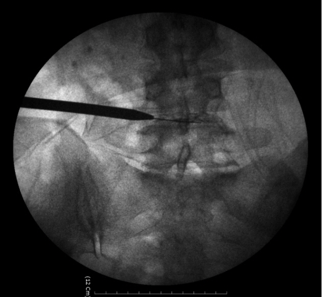
Fig. 3 Guide wire and obturator insertion.
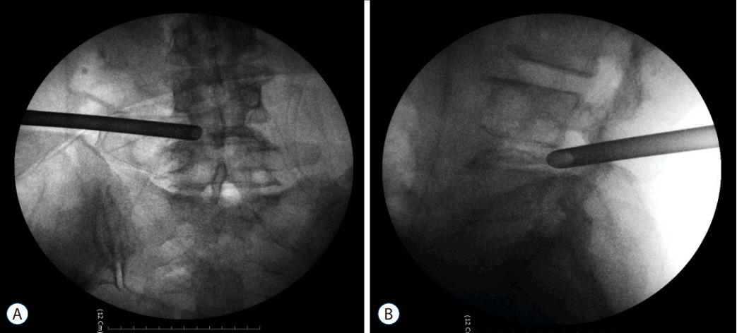
Fig. 4 Cannula positioning (A) anteroposterior and (B) lateral view.

Fig. 5 (A and B) Endoscopic view.

Fig. 6 Preoperative MRI. (A) Sagittal and (B) axial view. MRI: magnetic resonance imaging.
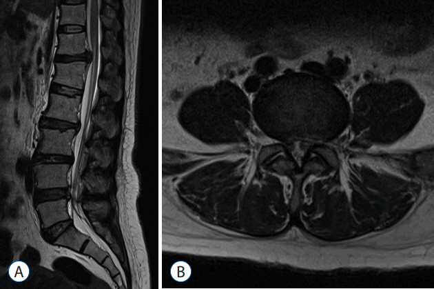
Fig. 7 Post-operative MRI. (A) Sagittal view and (B) axial view. MRI: magnetic resonance imaging.
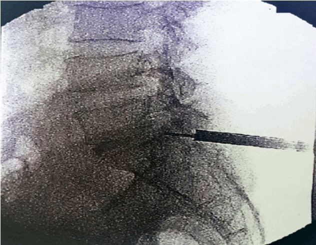
Fig. 8 C-arm positioning of cannula.
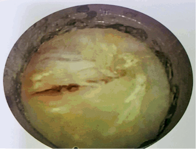
Fig. 9 Identification of two layers of the ligamentum flavum.
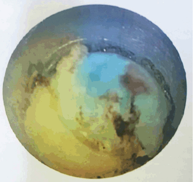
Fig. 10 Identification of Epidural fat and movements disc herniation.
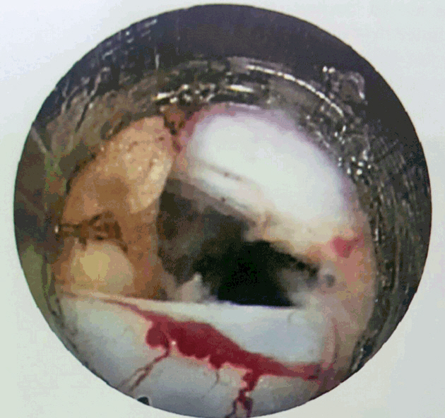
Fig. 11 End of decompression (free of the nerve roots).
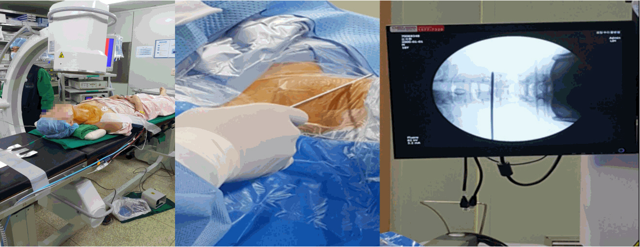
Fig. 12 Patient and C-arm positioning with level marking.

Fig. 13 Needle insertion.

Fig. 14 Discography.
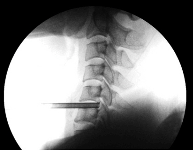
Fig. 15 Final position of cannula.

Fig. 16 Endoscope insertion.
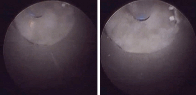
Fig. 17 Use of Ho: YAG laser to make annular window.

Fig. 18 End of decompression.
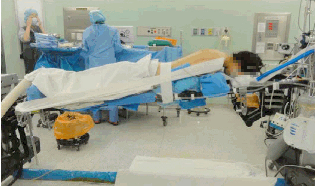
Fig. 19 Patient positioning.
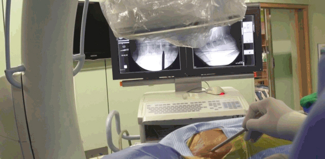
Fig. 20 Level marking.
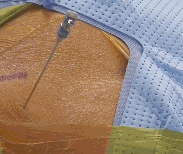
Fig. 21 Needle insertion.
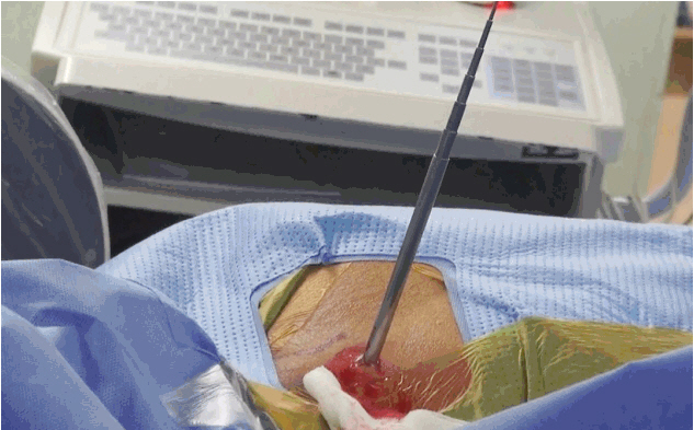
Fig. 22 Passage of serial dilators.
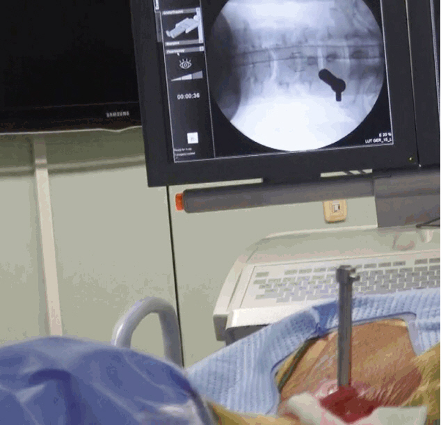
Fig. 23 Final positioning of cannula.
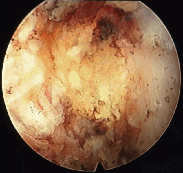
Fig. 24 Endoscopic view of facet (‘V’ shape area).

Fig. 25 Use of burr and kerrison punch for decompression.

Fig. 26 Use of burr and kerrison punch for decompression.
Fig. 27 Showing endoscopic approach to the thoracic spine.
Reference
-
References
1. Choi G, Lee SH, Raiturker PP, Lee S, Chae YS. Percutaneous endoscopic interlaminar discectomy for intracanalicular disc herniations at L5-S1 using a rigid working channel endoscope. Neurosurgery. 58(1 Suppl):ONS59–ONS68. discussion ONS59–ONS68. 2006.
Article2. Choi KY, Eun SS, Lee SH, Lee HY. Percutaneous endoscopic thoracic discectomy; transforaminal approach. Minim Invasive Neurosurg. 53:25–28. 2010.
Article3. Destandau J. A special device for endoscopic surgery of lumbar disc herniation. Neurol Res. 21:39–42. 1999.
Article4. Friedman WA. Percutaneous discectomy: an alternative to chemonucleolysis. J Neurosurg. 13:542–7. 1983.
Article5. Gore SR, Yeung AT. Identifying sources of discogenic pain. Jour Minimally Invasive Spinal Technique. 3:21–24. 2003.6. Hijikata S, Yamagishi M, Nakayama T, Oomori K. Percutaneous diskectomy: a new treatment method for lumbar disc herniation. J Toden Hosp. 5:5–13. 1975.7. Hoogland T. Transforaminal endoscopic discectomy with foraminoplasty for lumbar disc herniation. Surg Tech Orthop Traumatol. 40:55–120. 2003.8. Jho HD. Endoscopic transpedicular thoracic discectomy. J Neurosurg. 91(2 Suppl):151–156. 1999.
Article9. Kambin P. Arthroscopic microdiscectomy. Arthroscopy. 8:287–295. 1992.
Article10. Kambin P. Arthroscopic microdiscectomy. Kambin PR, editor. Minimal Intervention in Spinal Surgery. Baltimore: Urban & Schwarzenberg;1991. p. 67–1100.
Article11. Kambin P. Endoscopic laser discectomy. FDA;IDE G890238-S1. 1990.12. Kambin P. History of Surgical management of herniated lumbar discs from cauterization to arthroscopic and endoscopic spinal surgery. Kambin P, editor. Arthroscopic and Endoscopic Spinal Surgery. ed 2. Totowa: Humana Press Inc;2005. p. 1–27.
Article13. Kambin P, Gellman H. Percutaneous lateral discectomy of the lumbar spine: a preliminary report. Clin Orthop. 174:127–132. 1983.14. Kambin P, Sampson S. Posterolateral percutaneous suction-excision of herniated lumbar intervertebral discs. Report of interim results. Clin Orthop. (207):37–43. 1986.15. Kim DH, Choi G, Lee SH. Endoscopic spine proceduresed. New York: Thieme;2011. p. 42–58.16. Mathews HH. Transforaminal endoscopic microdiscectomy. Neurosurg Clin North Am. 7:59–63. 1996.
Article17. Mirkovic SR, Schwartz DG, Glazier KD. Anatomic considerations in posterolateral procedures. Spine (Phila Pa 1976). 20:1965–1971. 1995.18. Onik G, Helms CA, Ginsburg L, Hoaglund FT, Morris J. Percutaneous lumbar discectomy using new aspiration probe. AJR Am J Roentgenol. 144:1137–1140. 1985.19. Ruetten S, Komp M, Godolias G. An extreme lateral access for the surgery of lumbar disc herniations inside the spinal canal using the full-endoscopic uniportal transforaminal approach-technique and prospective results of 463 patients. Spine (Phila Pa 1976). 30:2570–2578. 2005.
Article20. Smith MM, Foley KT. Microendoscopic discectomy(MED): the first 100 cases. Neurosurgery. 43:702. 1998.21. Yeung AT. The evolution and advancement of endoscopic foraminal surgery: one surgeon’s experience incorporating adjunctive techologies. SAS J. 1:108–117. 2007.
Article22. Yeung AT, Gore SR. Evolving methodology in treating discogenic back pain by Selective Endoscopic Discectomy (SED) and thermal annuloplasty. J Minimally Invasive Spinal Tech. 1:8–16. 2001.
- Full Text Links
-
- Actions
-
Cited
- CITED
-
- Close
- Share
-
- Similar articles
-
- A Narrative Review of Development of Full-Endoscopic Lumbar Spine Surgery
- Complications and Management of Endoscopic Spinal Surgery
- The Role and Future of Endoscopic Spine Surgery: A Narrative Review
- Endoscopic Spine Surgery: Current State of Art and the Future Perspective
- Evolution of Spinal Endoscopic Surgery
