Three-dimensional EchoNavigator System Guided Transcatheter Closure of Paravalvular Leakage
- Affiliations
-
- 1Division of Cardiology, Department of Internal Medicine, Seoul St. Mary's Hospital, College of Medicine, The Catholic University of Korea, Seoul, Korea. peace816@catholic.ac.kr
- 2Division of Cardiology, Department of Internal Medicine, Incheon St. Mary's Hospital, College of Medicine, The Catholic University of Korea, Seoul, Korea.
- KMID: 2456859
- DOI: http://doi.org/10.4250/jcvi.2019.27.e30
Abstract
- No abstract available.
MeSH Terms
Figure
-
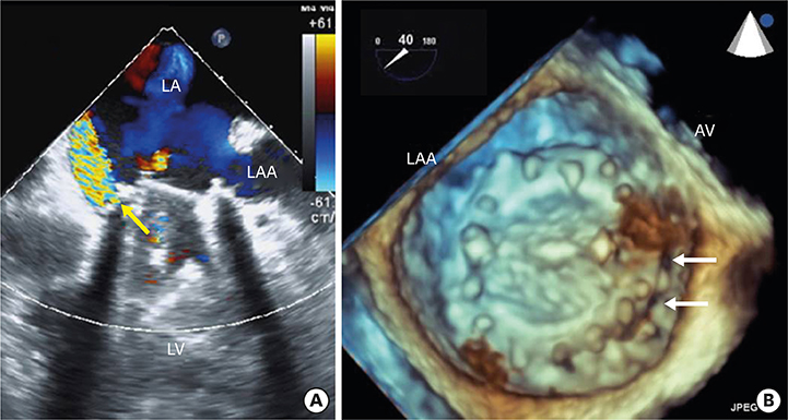
Figure 1 Pre-procedural transesophageal echocardiography (TEE). (A) Two-dimensional TEE color Doppler image demonstrated severe paravalvular leakage (yellow arrow). (B) Three-dimensional TEE image demonstrated dark holes between the prosthetic mitral valve and mitral annulus (white arrows), which was localized at the medial side of the anterior mitral annulus, near to the aortic valve. AV: aortic valve, LA: left atrium, LAA: left atrial appendage, LV: left ventricle.

Figure 2 Fusion image from the EchoNavigator System (Phillips Healthcare, Best, The Netherlands) during the procedure. Green dot, demonstrated on fluoroscopic view (A), was marked from the real-time 3-dimenstion transesophageal echocardiography image (B). The guide wire was targeted to the green dot on fluoroscopic view and was successfully passed through the slit.
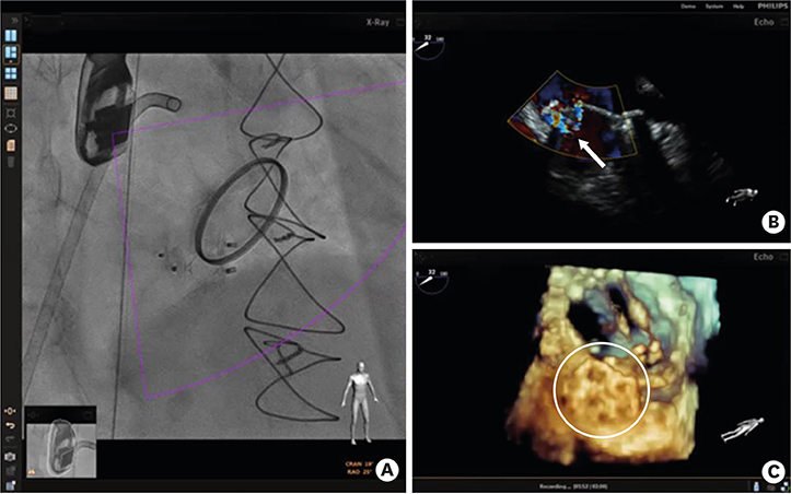
Figure 3 Fusion image from EchoNavigator System (Phillips Healthcare, Best, The Netherlands) after deployment. (A) Final positions of two Amplatzer™ vascular plugs are shown by fluoroscopy. (B) Color Doppler image demonstrated markedly reduced paravalvular leakage (white arrow) compared to Figure 1A. (C) Three-dimensional transesophageal echocardiography demonstrated final position of the vascular plugs (white circle).
Reference
-
1. Cruz-Gonzalez I, Rama-Merchan JC, Rodríguez-Collado J, et al. Transcatheter closure of paravalvular leaks: state of the art. Neth Heart J. 2017; 25:116–124.
Article2. Hascoet S, Smolka G, Bagate F, et al. Multimodality imaging guidance for percutaneous paravalvular leak closure: Insights from the multi-centre FFPP register. Arch Cardiovasc Dis. 2018; 111:421–431.
Article
- Full Text Links
-
- Actions
-
Cited
- CITED
-
- Close
- Share
-
- Similar articles
-
- Management of Recurrent Paravalvular Leakage in a Very High-Risk Patient: A Case Report
- Transcatheter Closures for Fistula Tract and Paravalvular Leak after Mitral Valve Replacement and Tricuspid Annuloplasty
- Comprehensive understanding of atrial septal defects by imaging studies for successful transcatheter closure
- Trido Mitral Valve Replacement with Dacron Collar Prosthetic Valve due to Paravalvular Leak
- Recurrent Paravalvular Leakage after Mitral Valve Replacement with Annular Reconstruction for Paravalvular Leakage Due to a Paravalvular Abscess: A case report
