Cotyledonoid dissecting leiomyoma: an uncommon form of a common disease
- Affiliations
-
- 1Department of Pathology, All India Institute of Medical Sciences, Phulwari Sharif, Patna, Bihar, India. rakesh.newjobi@gmail.com
- KMID: 2456833
- DOI: http://doi.org/10.5468/ogs.2019.62.5.362
Abstract
- Leiomyomas are benign uterine smooth muscle neoplasms with varied morphology that are well known to undergo secondary changes. Cotyledonoid dissecting leiomyoma is a rare and distinct form of leiomyoma that poses a diagnostic challenge for clinicians, radiologists, and pathologists and can be confused with malignant uterine neoplasms. Only a few cases have been reported so far in the literature. Here we report a case of a cotyledonoid dissecting leiomyoma in a 60-year-old woman, emphasize its gross and histological features, and provide a review of the literature.
Keyword
Figure
-
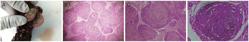
Fig. 1 Gross and histopathology images of cotyledonoid dissecting leiomyoma. (A) Cut section with multiple tan white nodules. (B) Classical whorling pattern (hematoxylin and eosin [HE], ×20). (C) Nodules of varying sizes of uniform smooth muscles arranged in interlacing and whorling fascicles with few prominent blood vessels (HE, ×100). (D) Bland smooth muscles arranged in an interlacing pattern with no signs of nuclear atypia, mitosis, or necrosis (HE, ×400).

Fig. 2 Immunohistochemistry images of cotyledonoid dissecting leiomyoma showing: (A) smooth muscle actin (immunohistochemistry [IHC], ×400); (B) Vimentin (IHC, ×400); and (C) Desmin positivity in smooth muscle fibers (IHC, ×400).

Fig. 3 Histopathological images of intramural leiomyoma showing: (A) Benign smooth muscles arranged in an interlacing pattern with large areas of hyalinization (hematoxylin and eosin [HE], ×100) and myometrium; and (B) Features of adenomyosis (HE, ×40).
Reference
-
1. Kim MJ, Park YK, Cho JH. Cotyledonoid dissecting leiomyoma of the uterus: a case report and review of the literature. J Korean Med Sci. 2002; 17:840–844.
Article2. Weissferdt A, Maheshwari MB, Downey GP, Rollason TP, Ganesan R. Cotyledonoid dissecting leiomyoma of the uterus: a case report. Diagn Pathol. 2007; 2:18.
Article3. Roth LM, Reed RJ, Sternberg WH. Cotyledonoid dissecting leiomyoma of the uterus. The Sternberg tumor. Am J Surg Pathol. 1996; 20:1455–1461.4. Menolascino-Bratta F, García de Barriola V, Naranjo de Gómez M, García Tamayo J, Suarez JA, Hernández Chacón AV. Cotyledonoid dissecting leiomyoma (Sternberg tumor): an unusual form of leiomyoma. Pathol Res Pract. 1999; 195:435–438.
Article5. Smith CC, Gold MA, Wile G, Fadare O. Cotyledonoid dissecting leiomyoma of the uterus: a review of clinical, pathological, and radiological features. Int J Surg Pathol. 2012; 20:330–341.6. Cramer SF, Patel A. The frequency of uterine leiomyomas. Am J Clin Pathol. 1990; 94:435–438.
Article7. Tanaka H, Toriyabe K, Senda T, Sakakura Y, Yoshida K, Asakura T, et al. Cotyledonoid dissecting leiomyoma treated by laparoscopic surgery: a case report. Asian J Endosc Surg. 2013; 6:122–125.
Article8. Anderson MC, Robboy SJ, Russell P. Chapter 15. Uterine smooth muscletumours. Pathology of the female reproductive tract. 1st ed. London: Churchill Livingstone;2002. p. 389–414.9. Gurbuz A, Karateke A, Kabaca C, Arik H, Bilgic R. A case of cotyledonoid leiomyoma and review of the literature. Int J Gynecol Cancer. 2005; 15:1218–1221.
Article
- Full Text Links
-
- Actions
-
Cited
- CITED
-
- Close
- Share
-
- Similar articles
-
- Uterine Leiomyomas with Perinodular Hydropic Degeneration: A Report of Two Cases
- Cotyledonoid Dissecting Leiomyoma of the Uterus with Intravascular Luminal Growth: A Case Study
- Cotyledonoid Dissecting Leiomyoma of the Uterus: A Case Report and Review of the Literature
- Uterine leiomyoma research
- A Case of Cutaneous Leiomyoma Presenting as a Large Nipple
