Importance of Magnetic Resonance Imaging Examination in Unossified Avulsion Fracture of Child Elbow
- Affiliations
-
- 1Department of Orthopedic Surgery, College of Medicine, Konyang University, Daejeon, Korea. ktk1113@hanmail.net
- KMID: 2456248
- DOI: http://doi.org/10.12790/ahm.2019.24.3.243
Abstract
- Unossified elbow avulsion fractures of children should be treated with care. Simple avulsion fractures seen on an x-ray may actually be severe injuries such as elbow joint subluxation or radial head dislocation. So, definite diagnosis is important in determining appropriate treatment of avulsion fractures around the unossified elbow, and magnetic resonance imaging (MRI) examination plays a key role in the diagnosis. We experienced avulsion fracture of the capitulum with common extensor tendon and lateral collateral ligament injuries and ulnar coronoid process fracture in an 11-year-old boy and radial head dislocation with common flexor tendon and partial ulnar collateral ligament injuries in an 8-year-old boy. We would like to talk about the importance of MRI in unossified avulsion fracture of child elbow by reviewing 2 case reports.
Keyword
MeSH Terms
Figure
-
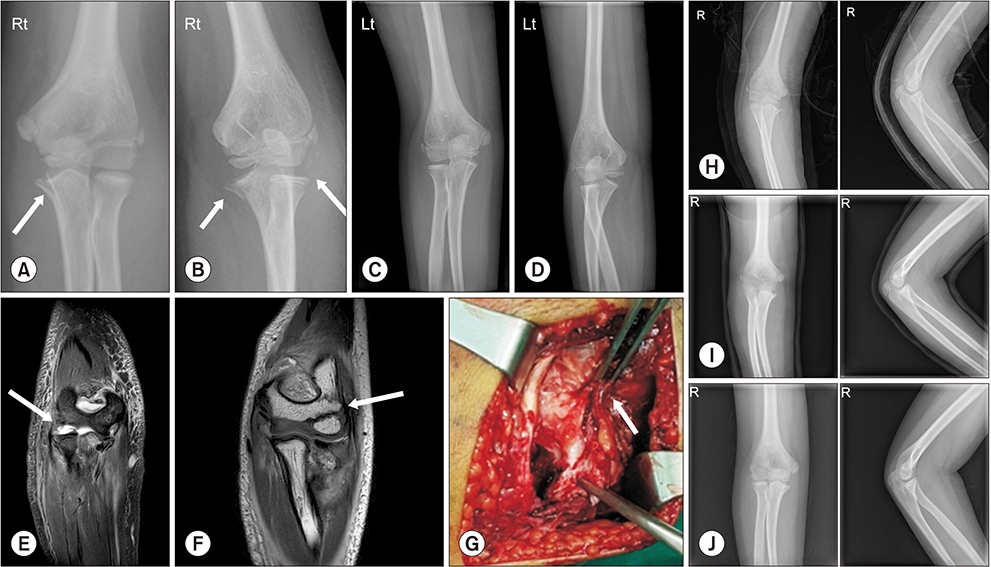
Fig. 1 Initial (A) anteroposterior and (B) oblique plain radiographs showed a right ulnar coronoid process fracture (arrows in A and B) and (C) anteroposterior and (D) oblique plain radiographs show the opposite side. (E) Magnetic resonance imaging showed radial head subluxation (arrow) with lateral collateral ligament injury. (F) Magnetic resonance imaging showed capitulum fracture (arrow) with common extensor tendon and collateral ligament injuries. (G) In gross photo, common extensor tendon and lateral collateral ligament were injured. (H), (I), and (J) plain radiographs taken after operation and in one, four month, respectively.
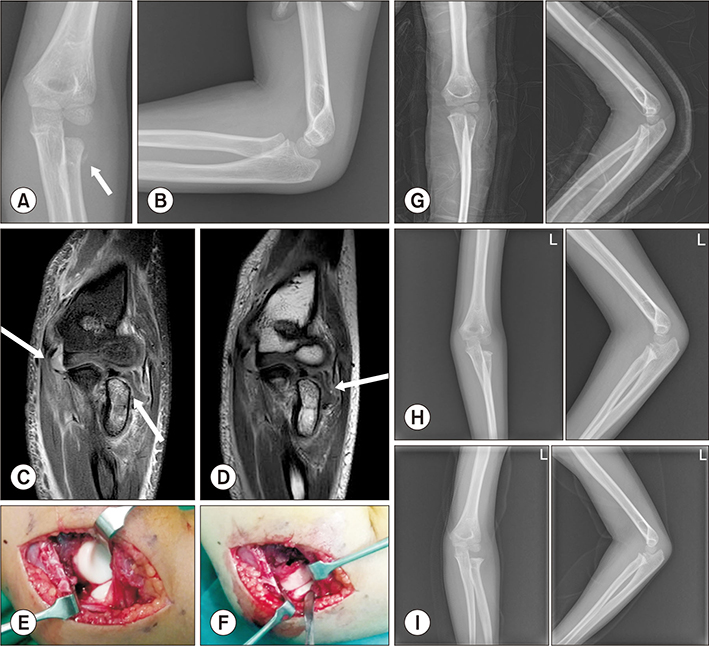
Fig. 2 Initial (A) anteroposterior (arrow) and (B) lateral radiograph of left elbow showed a bony fragment around the radial head. (C) and (D) magnetic resonance imaging showed a fracture with epiphyseal separation of radial head and neck (arrow at right lower in C and D) along with a partial tear of ulnar collateral ligament (arrow at left upper in C). (E) Radial head was completely dislocated and (F) reduced. (G) In postoperative radiograph, dislocated bony fragment was located on arrow. (H) After 1 month from operation, plan radiograph was taken. (I) After 5 months from operation, plan radiograph was taken.
Reference
-
1. Chessare JW, Rogers LF, White H, Tachdjian MO. Injuries of the medial epicondylar ossification center of the humerus. AJR Am J Roentgenol. 1977; 129:49–55.
Article2. Woods GW, Tullos HS. Elbow instability and medial epicondyle fractures. Am J Sports Med. 1977; 5:23–30.
Article3. Kamath AF, Baldwin K, Horneff J, Hosalkar HS. Operative versus non-operative management of pediatric medial epicondyle fractures: a systematic review. J Child Orthop. 2009; 3:345–357.
Article4. Miyazaki CS, Maranho DA, Agnollitto PM, Nogueira-Barbosa MH. Study of secondary ossification centers of the elbow in the Brazilian population. Acta Ortop Bras. 2017; 25:279–282.
Article5. Fowles JV, Slimane N, Kassab MT. Elbow dislocation with avulsion of the medial humeral epicondyle. J Bone Joint Surg Br. 1990; 72:102–104.
Article6. Tanabe K, Miyamoto N. Fracture of an unossified humeral medial epicondyle: use of magnetic resonance imaging for diagnosis. Skeletal Radiol. 2016; 45:1409–1412.
Article7. Pudas T, Hurme T, Mattila K, Svedström E. Magnetic resonance imaging in pediatric elbow fractures. Acta Radiol. 2005; 46:636–644.
Article8. Beltran J, Rosenberg ZS, Kawelblum M, Montes L, Bergman AG, Strongwater A. Pediatric elbow fractures: MRI evaluation. Skeletal Radiol. 1994; 23:277–281.
Article
- Full Text Links
-
- Actions
-
Cited
- CITED
-
- Close
- Share
-
- Similar articles
-
- Sleeve Fracture of the Superior Pole of Patella in an Adolescent
- MR Imaging of Lumbar Root Avulsion: Report of Two Case Studies
- Isolated Avulsion Fracture of the Subscapularis from the Lesser Tuberosity of the Humerus in a 12-Year-Old Boy - A Case Report -
- Pediatric Cartilaginous Tibia Eminence Fracture Overlooked on Plain Radiograph: A Report of Two Cases
- Intra-Apophyseal Avulsion Fracture of Medial Epicondyle in a Adolescent Baseball Player
