J Korean Soc Radiol.
2011 Jun;64(6):525-529. 10.3348/jksr.2011.64.6.525.
MR Imaging of Lumbar Root Avulsion: Report of Two Case Studies
- Affiliations
-
- 1Department of Radiology, Soonchunhyang University Hospital, Bucheon, Gyunggi-do, Korea. mj4907@schmc.ac.kr
- 2Department of Neurosurgery, Soonchunhyang University Hospital, Bucheon, Gyunggi-do, Korea.
- KMID: 1443500
- DOI: http://doi.org/10.3348/jksr.2011.64.6.525
Abstract
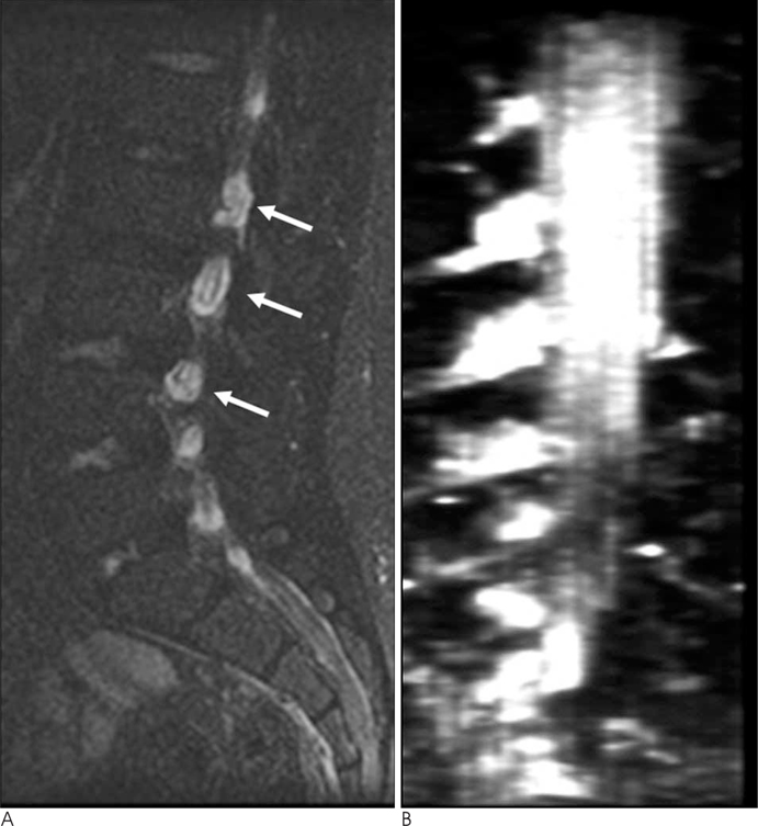
- Lumbosacral nerve root avulsion is a rare disease entity that is usually associated with a fracture of the pelvic bone. We report on two cases to share our multimodal images and to introduce new magnetic resonance (MR) imaging findings. In one case (a pediatric patient), neither a history of pelvic ring fracture, nor a history of hip dislocation with characteristic MR myelography findings was evident. In the other case, secondary MR findings associated with lumbosacral nerve root avulsion were found that had not been introduced previously.
MeSH Terms
Figure
Reference
-
1. Sasaka KK, Phisitkul P, Boyd JL, Marsh JL, El-Khoury GY. Lumbosacral nerve root avulsions: MR imaging demonstration of acute abnormalities. AJNR Am J Neuroradiol. 2006; 27:1944–1946.2. Foley JA, Rao RD. Traumatic lumbosacral pseudomeningocele associated with spinal fracture. Spine J. 2009; 9:E5–E10.3. Yoshikawa T, Hayashi N, Yamamoto S, Tajiri Y, Yoshioka N, Masumoto T, et al. Brachial plexus injury: clinical manifestations, conventional imaging findings, and the latest imaging techniques. Radiographics. 2006; 26:Suppl 1. S133–S143.4. Moschilla G, Song S, Chakera T. Post-traumatic lumbar nerve root avulsion. Australas Radiol. 2001; 45:281–284.5. Gasparotti R, Ferraresi S, Pinelli L, Crispino M, Pavia M, Bonetti M, et al. Three-dimensional MR myelography of traumatic injuries of the brachial plexus. AJNR Am J Neuroradiol. 1997; 18:1733–1742.6. Hans FJ, Reinges MH, Krings T. Lumbar nerve root avulsion following trauma: balanced fast field-echo MRI. Neuroradiology. 2004; 46:144–147.7. Cronin CG, Lohan DG, Meehan CP, Delappe E, McLoughlin R, O'Sullivan GJ, et al. Anatomy, pathology, imaging and intervention of the iliopsoas muscle revisited. Emerg Radiol. 2008; 15:295–310.8. Rozen WM, Tran TM, Ashton MW, Barrington MJ, Ivanusic JJ, Taylor GI. Refining the course of the thoracolumbar nerves: a new understanding of the innervation of the anterior abdominal wall. Clin Anat. 2008; 21:325–333.9. Parry GJ, Floberg J. Diabetic truncal neuropathy presenting as abdominal hernia. Neurology. 1989; 39:1488–1490.10. Billet FP, Ponssen H, Veenhuizen D. Unilateral paresis of the abdominal wall: a radicular syndrome caused by herniation of the L1-2 disc. J Neurol Neurosurg Psychiatry. 1989; 52:678.
- Full Text Links
- Actions
-
Cited
- CITED
-
- Close
- Share
- Similar articles
-
- MR Imaging of Traumatic Brachial Plexus Injury
- Avulsion Fracture of the Anterior Medial Meniscus Root
- Traumatic cervical root injury: Diagnostic value of MR imaging
- MR Imaging Findings of Avulsion Fracture of the Tibial Spine of the Knee, Focusing on Cruciate Ligament Tear
- Clinical Significance of Nerve Root Enhancement in Contrast-Enhanced MR Imaging of the Postoperative Lumbar Spine


