Ann Hepatobiliary Pancreat Surg.
2019 Aug;23(3):278-281. 10.14701/ahbps.2019.23.3.278.
Acalculous cholecystitis associated with Hantaan virus: A case report
- Affiliations
-
- 1Department of Surgery, Armed Forces Capital Hospital, Seongnam, Korea. dark8262@naver.com
- KMID: 2456027
- DOI: http://doi.org/10.14701/ahbps.2019.23.3.278
Abstract
- Acute acalculous cholecystitis (AAC) still remains one of the most elusive diagnoses and occurs in various conditions. Although AACs caused by viral infections are rare, various viruses have been revealed to cause AAC. Here we present a case in which a man suffered from AAC caused by a Hantaan virus infection. A 35-year-old man was referred to the emergency room for myalgia and fever that began 4 days ago. He suffered oliguria and abdominal pain for 2 days. At the time of his visit to the emergency room, he experienced a fever that spiked up to 38.3℃. An initial blood sample objectified the following pathologic results: white blood cells - 10260/µl; C-reactive protein - 6.76 mg/dl; total bilirubin - 1.7 mg/dl; AST - 90 IU/L; ALT - 233 IU/L. In the computed tomography, bilateral perirenal fluid collections and bilateral flexural effusion were shown and acute hepatopathy and cholecystopathy were also shown. Because there was no definite tenderness around the patient's right upper quadrant from physical examination and his cholecystopathy looked like it was from secondary change according to acute hepatopathy, we decided to perform conservative care without surgical treatment. The following day, in viral antibody test, Hantaan virus antibody was detected. After conservative management, the patient's condition improved and his laboratory findings were stable. The patient was discharged on the 10(th) day at the hospital stay without any symptoms. The Hantaan virus infection should be suspected as a causative agent of AAC, especially when there is abnormal liver function tests and abdominal pain.
Keyword
MeSH Terms
-
Abdominal Pain
Acalculous Cholecystitis*
Adult
Bilirubin
C-Reactive Protein
Cholecystitis
Diagnosis
Emergency Service, Hospital
Fever
Hantaan virus*
Hantavirus
Hemorrhagic Fever with Renal Syndrome
Humans
Length of Stay
Leukocytes
Liver Function Tests
Myalgia
Oliguria
Patient Rights
Physical Examination
Bilirubin
C-Reactive Protein
Figure
-
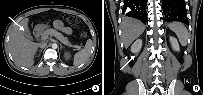
Fig. 1 Computed tomographic findings of the patient. (A) White arrow: pericholecystic fluid and subserosal edema with halo sign. (B) White wedge: pleural effusion, white arrow: perirenal fluid collections.
Reference
-
1. Barie PS, Eachempati SR. Acute acalculous cholecystitis. Gastroenterol Clin North Am. 2010; 39:343–357. x
Article2. Huffman JL, Schenker S. Acute acalculous cholecystitis: a review. Clin Gastroenterol Hepatol. 2010; 8:15–22.
Article3. Fröhlich R, Römmele U. [Acalculous cholecystitis in hantavirus infections]. Dtsch Med Wochenschr. 2013; 138:1255–1258. German.4. Keyaerts E, Ghijsels E, Lemey P, Maes P, Zachée P, Daelemans R, et al. Plasma exchange-associated immunoglobulin m-negative hantavirus disease after a camping holiday in southern france. Clin Infect Dis. 2004; 38:1350–1356.
Article5. Nicolas JB. Acalculous cholecystitis associated with hemorrhagic fever with renal syndrome. Acta Clin Belg. 2015; 70:377–381.
Article6. Lee HW, Lee PW, Johnson KM. Isolation of the etiologic agent of Korean hemorrhagic fever. 1978. J Infect Dis. 2004; 190:1711–1721.7. Avšič-Županc T, Saksida A, Korva M. Hantavirus infections. Clin Microbiol Infect. 2019; 21S:e6–e16.
Article8. Mackow ER, Gavrilovskaya IN. Hantavirus regulation of endothelial cell functions. Thromb Haemost. 2009; 102:1030–1041.
Article9. Kim YO, Chun KA, Choi JY, Yoon SA, Yang CW, Kim KT, et al. Sonographic evaluation of gallbladder-wall thickening in hemorrhagic fever with renal syndrome: prediction of disease severity. J Clin Ultrasound. 2001; 29:286–289.
Article10. Poddighe D, Sazonov V. Acute acalculous cholecystitis in children. World J Gastroenterol. 2018; 24:4870–4879.
Article
- Full Text Links
-
- Actions
-
Cited
- CITED
-
- Close
- Share
-
- Similar articles
-
- A Case of Spontaneous Hemorrhagic Cholecystitis without Gallstone
- A Case of Hemobilia Caused by Chronic Acalculous Cholecystitis
- Acalculous Hemorrhagic Cholecystitis with Chronic Intraluminal Hematoma: MRI Findings
- Acute acalculous cholecystitis
- A Case of Churg-Strauss Syndrome Which First Presented as Acute Cholecystitis
