Korean J Radiol.
2019 Sep;20(9):1390-1398. 10.3348/kjr.2018.0914.
De Novo Intracranial Aneurysms Detected on Imaging Follow-Up of Coiled Aneurysms in a Korean Population
- Affiliations
-
- 1Department of Radiology, KyungHee University Medical Center, KyungHee University College of Medicine, Seoul, Korea.
- 2Department of Radiology, Seoul National University Hospital, Seoul National University College of Medicine, Seoul, Korea. aronnn@naver.com
- 3Department of Neurosurgery, Dongguk University Ilsan Hospital, Dongguk University College of Medicine, Goyang, Korea.
- 4Department of Neurosurgery, Seoul National University Hospital, Seoul National University College of Medicine, Seoul, Korea.
- 5Department of Radiology, Veterans Health Service Medical Center, Seoul, Korea.
- KMID: 2455767
- DOI: http://doi.org/10.3348/kjr.2018.0914
Abstract
OBJECTIVE
Coiled aneurysms are known to recanalize over time, making follow-up evaluations mandatory. Although de novo intracranial aneurysms (DNIAs) are occasionally detected during routine patient monitoring, such events have not been thoroughly investigated to date. Herein, we generated estimates of DNIA development during long-term observation of coiled cerebral aneurysms, focusing on incidence and the risk factors involved.
MATERIALS AND METHODS
In total, 773 patients undergoing coil embolization of intracranial aneurysms between 2008 and 2010 were reviewed retrospectively. Their medical records and radiologic data accrued over the extended period (mean, 52.7 ± 29.7 months) were analyzed. For the detection of DNIA, follow-up magnetic resonance angiography and/or conventional angiography were used. The incidence of DNIAs and related risk factors were analyzed using Cox proportional hazards regression and Kaplan-Meier product-limit estimator.
RESULTS
In 19 (2.5%) of the 773 patients with coiled aneurysms, DNIAs (0.56% per patient-year) developed during continued long-term monitoring (3395.3 patient-years). Of these, 9 DNIAs (47.4%) were detected within 60 months, with 10 (52.6%) emerging thereafter. The most common site involved was the posterior communicating artery (n = 6), followed by the middle cerebral artery (n = 5) and the basilar top (n = 4). Multivariate analysis indicated that younger age (< 50 years) (hazard ratio [HR] = 1.045; p = 0.010) and recanalization of coiled aneurysms (HR = 2.560; p = 0.047) were significant factors in DNIA formation, whereas female sex, smoking, and hypertension fell short of statistical significance. Cumulative survival rates without DNIA were significantly higher in older subjects (> 60 years; p < 0.001) and in the absence of post-coiling aneurysm recurrence (p = 0.006).
CONCLUSION
In most patients with coiled aneurysms, development of DNIAs during long-term monitoring is rare. However, younger patients (< 50 years) or patients with recurring aneurysms appear to be predisposed to DNIAs.
Keyword
MeSH Terms
-
Aneurysm*
Angiography
Arteries
Embolization, Therapeutic
Female
Follow-Up Studies*
Humans
Hypertension
Incidence
Intracranial Aneurysm*
Magnetic Resonance Angiography
Medical Records
Middle Cerebral Artery
Monitoring, Physiologic
Multivariate Analysis
Recurrence
Retrospective Studies
Risk Factors
Smoke
Smoking
Survival Rate
Smoke
Figure
-
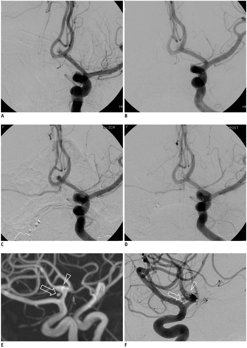
Fig. 1 Representative case of de novo aneurysm.(A) Pre- and (B) post-embolization angiographic images of ruptured anterior communicating artery aneurysm. C. Conventional angiography with 6-month follow-up shows major recanalization of coiled aneurysm. D. Post-procedural angiography after additional coiling confirms successful occlusion of aneurysm. (E) Magnetic resonance and (F) conventional angiography in 60-month follow-up show de novo aneurysm (arrows) adjacent to coiled aneurysm (arrowheads).
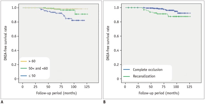
Fig. 2 Kaplan-Meier estimates of de novo aneurysm-free cumulative survival for (A) age at initial diagnosis of aneurysm and (B) recanalization of coiled aneurysm.DNIA = de novo intracranial aneurysm
Reference
-
1. Nieuwkamp DJ, Setz LE, Algra A, Linn FH, de Rooij NK, Rinkel GJ. Changes in case fatality of aneurysmal subarachnoid haemorrhage over time, according to age, sex, and region: a meta-analysis. Lancet Neurol. 2009; 8:635–642. PMID: 19501022.
Article2. Vlak MH, Algra A, Brandenburg R, Rinkel GJ. Prevalence of unruptured intracranial aneurysms, with emphasis on sex, age, comorbidity, country, and time period: a systematic review and meta-analysis. Lancet Neurol. 2011; 10:626–636. PMID: 21641282.
Article3. Giordan E, Lanzino G, Rangel-Castilla L, Murad MH, Brinjikji W. Risk of de novo aneurysm formation in patients with a prior diagnosis of ruptured or unruptured aneurysm: systematic review and meta-analysis. J Neurosurg. 2018; 131:1–11.
Article4. Ferns SP, Sprengers ME, van Rooij WJ, van den Berg R, Velthuis BK, de Kort GA, et al. De novo aneurysm formation and growth of untreated aneurysms: a 5-year MRA follow-up in a large cohort of patients with coiled aneurysms and review of the literature. Stroke. 2011; 42:313–318. PMID: 21164110.5. Lindgren AE, Räisänen S, Björkman J, Tattari H, Huttunen J, Huttunen T, et al. De novo aneurysm formation in carriers of saccular intracranial aneurysm disease in Eastern Finland. Stroke. 2016; 47:1213–1218. PMID: 27026632.
Article6. Rahmah NN, Horiuchi T, Kusano Y, Sasaki T, Hongo K. De novo aneurysm: case reports and literature review. Neurosurgery. 2011; 69:E761–E766. discussion E766–E767. PMID: 21471840.
Article7. Zali A, Khoshnood RJ, Zarghi A. De novo aneurysms in long-term follow-up computed tomographic angiography of patients with clipped intracranial aneurysms. World Neurosurg. 2014; 82:722–725. PMID: 23827320.
Article8. Kheireddin AS, Filatov YM, Belousova OB, Eliava SS, Sazonov IA, Kaftanov AN, et al. [De novo cerebral aneurysms]. Zh Vopr Neirokhir Im N N Burdenko. 2015; 79:75–81.
Article9. Miller CA, Hill SA, Hunt WE. “De novo” aneurysms. A clinical review. Surg Neurol. 1985; 24:173–180. PMID: 4012574.
Article10. Sprengers ME, van Rooij WJ, Sluzewski M, Rinkel GJ, Velthuis BK, de Kort GA, et al. MR angiography follow-up 5 years after coiling: frequency of new aneurysms and enlargement of untreated aneurysms. AJNR Am J Neuroradiol. 2009; 30:303–307. PMID: 18971290.
Article11. Kemp WJ 3rd, Fulkerson DH, Payner TD, Leipzig TJ, Horner TG, Palmer EL, et al. Risk of hemorrhage from de novo cerebral aneurysms. J Neurosurg. 2013; 118:58–62. PMID: 23061385.
Article12. Wang JY, Smith R, Ye X, Yang W, Caplan JM, Radvany MG, et al. Serial imaging surveillance for patients with a history of intracranial aneurysm: risk of de novo aneurysm formation. Neurosurgery. 2015; 77:32–42. discussion 42–43. PMID: 25790068.13. Serrone JC, Tackla RD, Gozal YM, Hanseman DJ, Gogela SL, Vuong SM, et al. Aneurysm growth and de novo aneurysms during aneurysm surveillance. J Neurosurg. 2016; 125:1374–1382. PMID: 26967775.
Article14. Juvela S, Poussa K, Porras M. Factors affecting formation and growth of intracranial aneurysms: a long-term follow-up study. Stroke. 2001; 32:485–491. PMID: 11157187.15. Wermer MJ, van der Schaaf IC, Velthuis BK, Algra A, Buskens E, Rinkel GJ. ASTRA Study Group. Follow-up screening after subarachnoid haemorrhage: frequency and determinants of new aneurysms and enlargement of existing aneurysms. Brain. 2005; 128(Pt 10):2421–2429. PMID: 16000333.
Article16. van der Schaaf IC, Velthuis BK, Wermer MJ, Majoie C, Witkamp T, de Kort G, et al. ASTRA Study Group. New detected aneurysms on follow-up screening in patients with previously clipped intracranial aneurysms: comparison with DSA or CTA at the time of SAH. Stroke. 2005; 36:1753–1758. PMID: 16002762.17. Burkhardt JK, Chua MHJ, Weiss M, Do ASS, Winkler EA, Lawton MT. Risk of aneurysm residual regrowth, recurrence, and de novo aneurysm formation after microsurgical clip occlusion based on follow-up with catheter angiography. World Neurosurg. 2017; 106:74–84. PMID: 28648910.
Article18. Bruneau M, Rynkowski M, Smida-Rynkowska K, Brotchi J, De Witte O, Lubicz B. Long-term follow-up survey reveals a high yield, up to 30% of patients presenting newly detected aneurysms more than 10 years after ruptured intracranial aneurysms clipping. Neurosurg Rev. 2011; 34:485–496. PMID: 21643681.19. Cho YD, Kim KM, Lee WJ, Sohn CH, Kang HS, Kim JE, et al. Time-of-flight magnetic resonance angiography for follow-up of coil embolization with enterprise stent for intracranial aneurysm: usefulness of source images. Korean J Radiol. 2014; 15:161–168. PMID: 24497808.
Article20. Roy D, Milot G, Raymond J. Endovascular treatment of unruptured aneurysms. Stroke. 2001; 32:1998–2004. PMID: 11546888.
Article21. TakahashiS. Neurovascular imaging: MRI & microangiography. London: Springer-Verlag;2010. p. 345–372.22. Jabbarli R, Dinger TF, Darkwah Oppong M, Pierscianek D, Dammann P, Wrede KH, et al. Risk factors for and clinical consequences of multiple intracranial aneurysms: a systematic review and meta-analysis. Stroke. 2018; 49:848–855. PMID: 29511128.23. David CA, Vishteh AG, Spetzler RF, Lemole M, Lawton MT, Partovi S. Late angiographic follow-up review of surgically treated aneurysms. J Neurosurg. 1999; 91:396–401. PMID: 10470813.
Article24. Kim ST, Jeong HW, Jeong YG, In HS. Coiling as retreatment in intracranial aneurysm of de novo formation or regrowth: case report. Neurointervention. 2013; 8:46–51. PMID: 23515648.25. Brown MA, Parish J, Guandique CF, Payner TD, Horner T, Leipzig T, et al. A long-term study of durability and risk factors for aneurysm recurrence after microsurgical clip ligation. J Neurosurg. 2017; 126:819–824. PMID: 27128583.
Article26. Bor AS, Rinkel GJ, van Norden J, Wermer MJ. Long-term, serial screening for intracranial aneurysms in individuals with a family history of aneurysmal subarachnoid haemorrhage: a cohort study. Lancet Neurol. 2014; 13:385–392. PMID: 24618352.
Article27. Miyazawa N, Akiyama I, Yamagata Z. Risk factors for growth of unruptured intracranial aneurysms: follow-up study by serial 0.5-T magnetic resonance angiography. Neurosurgery. 2006; 58:1047–1053. PMID: 16723883.
Article28. Juvela S, Poussa K, Lehto H, Porras M. Natural history of unruptured intracranial aneurysms: a long-term follow-up study. Stroke. 2013; 44:2414–2421. PMID: 23868274.
Article29. Lai LT, Morgan MK, Patel NJ. Smoking increases the risk of de novo intracranial aneurysms. World Neurosurg. 2014; 82:e195–e201. PMID: 24518886.
Article30. Phan TG, Huston J 3rd, Brown RD Jr, Wiebers DO, Piepgras DG. Intracranial saccular aneurysm enlargement determined using serial magnetic resonance angiography. J Neurosurg. 2002; 97:1023–1028. PMID: 12450022.
Article31. Goksu E, Korkmaz E, Akyuz M, Ozgur O, Sindel T, Tuncer R. The analysis of long-term follow-up screening in patients with surgically treated intracranial aneurysms. Turk Neurosurg. 2015; 25:404–409. PMID: 26037180.32. Akyüz M, Tuncer R, Yilmaz S, Sindel T. Angiographic follow-up after surgical treatment of intracranial aneurysms. Acta Neurochir (Wien). 2004; 146:245–250. discussion 250. PMID: 15015046.33. Lecler A, Raymond J, Rodriguez-Régent C, Al Shareef F, Trystram D, Godon-Hardy S, et al. Intracranial aneurysms: recurrences more than 10 years after endovascular treatment-a prospective cohort study, systematic review, and meta-analysis. Radiology. 2015; 277:173–180. PMID: 26057784.
Article34. Cho YD, Jeon JP, Yoo DH, Cho WS, Kang HS, Kim JE, et al. Growth-related major recanalization of coiled aneurysms: incidence and risk factors. Neurosurgery. 2018; 82:185–191. PMID: 28605492.
Article35. Abdihalim M, Watanabe M, Chaudhry SA, Jagadeesan B, Suri MF, Qureshi AI. Are coil compaction and aneurysmal growth two distinct etiologies leading to recurrence following endovascular treatment of intracranial aneurysm? J Neuroimaging. 2014; 24:171–175. PMID: 23317437.
Article36. Xu L, Sugawara M, Tanaka G, Ohta M, Liu H, Yamaguchi R. Effect of elasticity on wall shear stress inside cerebral aneurysm at anterior cerebral artery. Technol Health Care. 2016; 24:349–357. PMID: 26835728.
Article37. Lee CJ, Zhang Y, Takao H, Murayama Y, Qian Y. A fluid-structure interaction study using patient-specific ruptured and unruptured aneurysm: the effect of aneurysm morphology, hypertension and elasticity. J Biomech. 2013; 46:2402–2410. PMID: 23962529.
Article38. Jahed M, Ghalichi F, Farhoudi M. Fluid-structure interaction of patient-specific Circle of Willis with aneurysm: investigation of hemodynamic parameters. Biomed Mater Eng. 2018; 29:357–368. PMID: 29578465.
Article39. Molyneux AJ, Kerr RS, Birks J, Ramzi N, Yarnold J, Sneade M, et al. ISAT Collaborators. Risk of recurrent subarachnoid haemorrhage, death, or dependence and standardised mortality ratios after clipping or coiling of an intracranial aneurysm in the International Subarachnoid Aneurysm Trial (ISAT): long-term follow-up. Lancet Neurol. 2009; 8:427–443. PMID: 19329361.
Article
- Full Text Links
-
- Actions
-
Cited
- CITED
-
- Close
- Share
-
- Similar articles
-
- De novo Formation of Familial Cerebral Aneurysms
- Intracranial "De Novo" Aneurysms: Case Report
- Coiling as Retreatment in Intracranial Aneurysm of de novo Formation or Regrowth: Case Report
- Role of Three-dimensional Computed Tomography Angiography in the Follow-up of Patients with Aneurysm Clips
- Guideline for Management of Unruptured Intracranial Aneurysms: Preliminary Report
