J Korean Ophthalmol Soc.
2019 Aug;60(8):719-724. 10.3341/jkos.2019.60.8.719.
Short-term Clinical Outcomes of Small Incision Lenticule Extraction for Correction of Myopia Patients with Corneal Opacity
- Affiliations
-
- 1Onnuri Smile Eye Clinic, Seoul, Korea. ytchungc@daum.net
- KMID: 2455453
- DOI: http://doi.org/10.3341/jkos.2019.60.8.719
Abstract
- PURPOSE
To evaluate the clinical outcomes of small incision lenticule extraction (SMILE) for the treatment of myopia with corneal opacity.
METHODS
Thirteen eyes of 13 patients with corneal opacities who underwent SMILE were retrospectively studied. Uncorrected distance visual acuity, spherical equivalence, efficacy index, and safety index were noted at 3 months after the procedure. The density and area of the corneal opacities were measured and compared preoperatively and 3 months postoperatively.
RESULTS
All eyes had preoperative corneal opacities within the lenticule formation areas. The mean area and density of corneal opacities were 0.72 ± 0.33 mm2 and 52.46 ± 15.74, respectively. All procedures were uneventful and no intraoperative complications were observed. At 3 months after the procedure, the efficacy and safety indices were 1.01 ± 0.15, and 1.05 ± 0.10, respectively, and the mean densities and areas of corneal opacities were 46.85 ± 14.56 (p = 0.038) and 0.70 ± 0.36 mm2 (p = 0.776), respectively.
CONCLUSIONS
The SMILE procedure was effective and safe for the correction of myopic patients with corneal opacities.
MeSH Terms
Figure
-
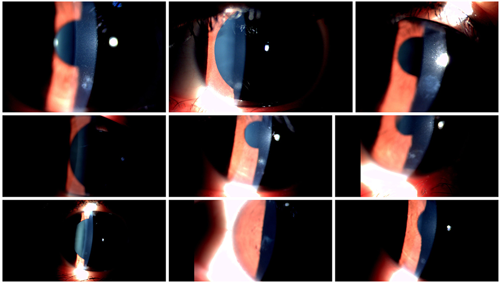
Figure 1 Anterior segment photos of case No. 2, 3, 4, 5, 6, 7, 9, 10, 12. The corneal opacity of all eyes was within the lenticule formation area.
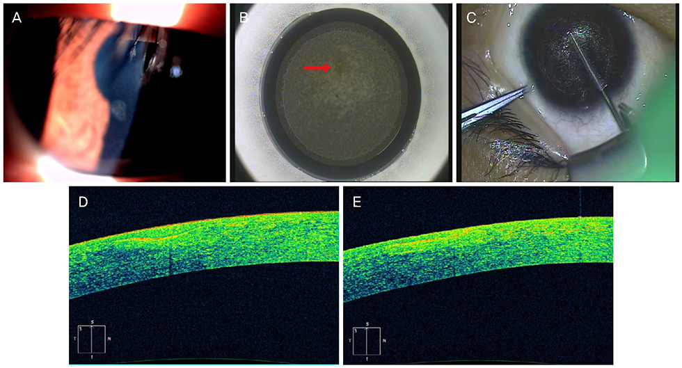
Figure 2 The preoperative, intraoperative, and postoperative corneal examinations in case No. 1. (A) The preoperative corneal opacity was noted by slit-lamp microscopy. (B) Corneal opacity was noted during lenticule scanning (red arrow). (C) The lenticule was separated successfully. (D) The preoperative anterior segment optical coherence tomography (AS-OCT) image. Thickening of epithelium and hyper-reflectivity of anterior stroma at corneal opacity lesion was noted. (E) The postoperative AS-OCT image. Thinning of the opacity lesion was noted.
Reference
-
1. Gupta N, Vashist P, Tandon R, et al. Prevalence of corneal diseases in the rural Indian population: the Corneal Opacity Rural Epidemiological (CORE) study. Br J Ophthalmol. 2015; 99:147–152.
Article2. Singh K, Jain D, Teli K. Rehabilitation of vision disabling corneal opacities: is there hope without corneal transplant? Cont Lens Anterior Eye. 2013; 36:74–79.
Article3. Soong HK, Malta JB. Femtosecond lasers in ophthalmology. Am J Ophthalmol. 2009; 147:189–197.
Article4. Vestergaard A, Ivarsen AR, Asp S, Hjortdal JØ. Small-incision lenticule extraction for moderate to high myopia: predictability, safety, and patient satisfaction. J Cataract Refract Surg. 2012; 38:2003–2010.
Article5. Miao H, Tian M, Xu Y, et al. Visual outcomes and optical quality after femtosecond laser small incision lenticule extraction: an 18-month prospective study. J Refract Surg. 2015; 31:726–731.
Article6. Blum M, Täubig K, Gruhn C, et al. Five-year results of small incision lenticule extraction (ReLEx SMILE). Br J Ophthalmol. 2016; 100:1192–1195.
Article7. Miao H, He L, Shen Y, et al. Optical quality and intraocular scattering after femtosecond laser small incision Lenticule extraction. J Refract Surg. 2014; 30:296–302.
Article8. Tomita M, Chiba A, Matsuda J, Nawa Y. Evaluation of LASIK treatment with the Femto LDV in patients with corneal opacity. J Refract Surg. 2012; 28:25–30.
Article9. Zhang S, Xu H, Zheng K, et al. The observation during small incision lenticule extraction for myopia with corneal opacity. BMC Ophthalmol. 2017; 17:80.
Article10. Prakash G, Avadhani K, Kalliath J, Srivastava D. Implantable collamer lens in a case of corneal scar with anisometropic amblyopia in an adult: an expanded indication. BMJ Case Rep. 2015; 2015:pii: bcr2014208862.
Article11. Barsam A, Allan BD. Excimer laser refractive surgery versus phakic intraocular lenses for the correction of moderate to high myopia. Cochrane Database Syst Rev. 2010; (5):CD007679.
Article12. Kamiya K, Shimizu K, Kobashi H, et al. Three-year follow-up of posterior chamber toric phakic intraocular lens implantation for the correction of high myopic astigmatism in eyes with keratoconus. Br J Ophthalmol. 2015; 99:177–183.
Article13. Farid M, Steinert RF. Femtosecond laser-assisted corneal surgery. Curr Opin Ophthalmol. 2010; 21:288–292.
Article14. Choi SK, Kim JH, Lee D. Treatment of corneal opacity by planned lamellar keratectomy using the femtosecond laser. Cornea. 2011; 30:907–909.
Article15. von Jagow B, Kohnen T. Corneal architecture of femtosecond laser and microkeratome flaps imaged by anterior segment optical coherence tomography. J Cataract Refract Surg. 2009; 35:35–41.
Article16. Ishikawa S, Kato N, Takeuchi M. Quantitative evaluation of corneal epithelial edema after cataract surgery using corneal densitometry: a prospective study. BMC Ophthalmol. 2018; 18:334.
Article17. Cennamo G, Forte R, Aufiero B, La Rana A. Computerized Scheimpflug densitometry as a measure of corneal optical density after excimer laser refractive surgery in myopic eyes. J Cataract Refract Surg. 2011; 37:1502–1506.
Article18. Rashad MA. Pentacam-based phototherapeutic keratectomy outcome in superficial corneal opacities. Clin Ophthalmol. 2012; 6:885–894.
Article
- Full Text Links
-
- Actions
-
Cited
- CITED
-
- Close
- Share
-
- Similar articles
-
- Clinical Outcomes of Small Incision Lenticule Extraction in Myopia: Study of Vector Parameters and Corneal Aberrations
- Clear lens Extraction and Epikeratophakic Lenticule Removal in Complicated Epikeratophakic Patients
- Outcomes of Small Incision Lenticule Extraction: Mild to Moderate Myopia versus High Myopia
- Comparison of Clinical Results of Lens Extraction and LASIK for Correction of High Myopia
- Comparison of Corneal Haze according to the Success of Corneal Epithelial Flap after Laser Subepithelial Keratomileusis
