MRI Findings of Accessory Popliteus Muscle: A Case Report
- Affiliations
-
- 1Department of Radiology, Inje University Seoul Paik Hospital, College of Medicine, Inje University, Seoul, Korea. shimage@naver.com
- KMID: 2454038
- DOI: http://doi.org/10.3348/jksr.2019.80.3.574
Abstract
- Accessory muscles located in the region of the popliteal fossa are very rare. MRI scan performed in a 52-year-old man with right knee pain revealed an anomalous muscle in the region of the popliteal fossa. Considering the muscle originated from the medial side of the lateral head of the gastrocnemius muscle and attached to the posteromedial articular capsule of the knee joint, it is consistent with the accessory popliteus muscle, previously reported. To our best knowledge, MRI finding about the accessory popliteus muscle has been reported in only one case. We present a case of the accessory popliteus muscle incidentally identified on MRI.
MeSH Terms
Figure
-
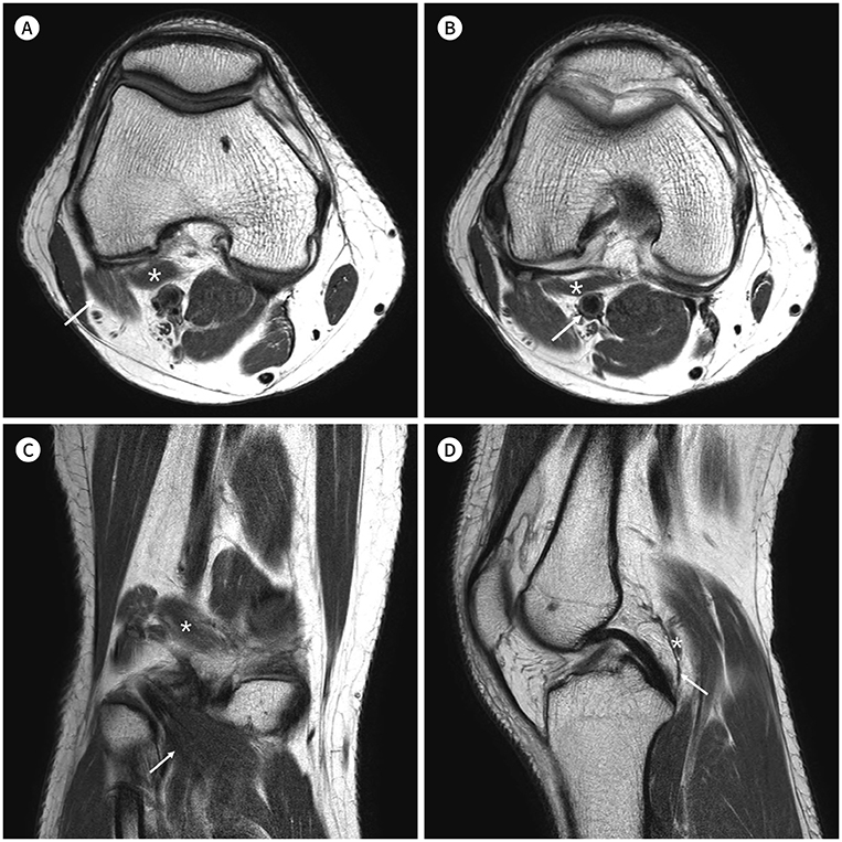
Fig. 1 MR images of the accessory popliteus muscle in a 52-year-old man who presented with right knee pain. A. Axial intermediate-weighted TSE image (TR/TE, 4000/36) shows the accessory popliteus muscle (asterisk) originating from the medial side of the lateral head of the gastrocnemius muscle (arrow). B. At 10.5 mm below the level of (A), the accessory popliteus muscle (asterisk) passes anterior to the popliteal artery (arrow) within the deep popliteal fossa. C. Coronal intermediate-weighted TSE image (TR/TE, 4000/36) demonstrates the accessory popliteal muscle (asterisk) coursing in an inferomedial direction, parallel to the normal popliteus muscle (arrow). D. Sagittal intermediate-weighted TSE image (TR/TE, 4000/36) shows the accessory popliteus muscle (asterisk) inserting into the posteromedial articular capsule (arrow). TE = echo time, TR = repetition time, TSE = turbo spin echo A B
Reference
-
1. Standring S. Grays's anatomy: the anatomical basis of clinical practice. 41th ed. New York: Elsevier;2016. p. 1397.2. Montet X, Sandoz A, Mauget D, Martinoli C, Bianchi S. Sonographic and MRI appearance of tensor fasciae suralis muscle, an uncommon cause of popliteal swelling. Skeletal Radiol. 2002; 31:536–538.
Article3. Stoane JM, Gordon DH. MRI of an accessory semimembranosus muscle. J Comput Assist Tomogr. 1995; 19:161–162.
Article4. Duc SR, Wentz KU, Käch KP, Zollikofer CL. First report of an accessory popliteal muscle: detection with MRI. Skeletal Radiol. 2004; 33:429–431.
Article5. Sobel M, Levy ME, Bohne WH. Congenital variations of the peroneus quartus muscle: an anatomic study. Foot Ankle. 1990; 11:81–89.
Article6. Lorentzon R, Wirell S. Anatomic variations of the accessory soleus muscle. Acta Radiol. 1987; 28:627–629.
Article7. Bowers CA, Mendicino RW, Catanzariti AR, Kernick ET. The flexor digitorum accessories longus-a cadaveric study. J Foot Ankle Surg. 2009; 48:111–115.8. Sookur PA, Naraghi AM, Bleakney RR, Jalan R, Chan O, White LM. Accessory muscles: anatomy, symptoms, and radiologic evaluation. Radiographics. 2008; 28:481–499.
Article9. Elias DA, White LM, Rubenstein JD, Christakis M, Merchant N. Clinical evaluation and MR imaging features of popliteal artery entrapment and cystic adventitial disease. AJR Am J Roentgenol. 2003; 180:627–632.
Article10. Bejjani FJ, Jahss MH. Le Double's study of muscle variations of the human body, part I: muscle variations of the leg. Foot Ankle. 1985; 6:111–134.
Article
- Full Text Links
-
- Actions
-
Cited
- CITED
-
- Close
- Share
-
- Similar articles
-
- The MRI Findings of Flexor Digitorum Accessorius Longus Muscle: a Case Report
- Accessory Hamstring Muscle: A Case Report
- Unilateral Accessory Cleidohyoid Muscle: A Case Report
- An accessory muscle of flexor digitorum profundus with bipennate first lumbrical: a unique variation of clinical significance
- Accessory Belly of the Piriformis Muscle as a Cause of Piriformis Syndrome: a Case Report with Magnetic Resonance Imaging and Magnetic Resonance Neurography Imaging Findings
