Ossification of the Triradiate Cartilage and Posterior Acetabulum
- Affiliations
-
- 1Department of Radiology, Kangwon National University Hospital, Chuncheon, Korea. hk2005.yoon@gmail.com
- KMID: 2454029
- DOI: http://doi.org/10.3348/jksr.2019.80.3.503
Abstract
- PURPOSE
This study attempts to evaluate the skeletal maturation patterns of the triradiate cartilage (TRC) and the posterior acetabular wall (PA), which can be easily assessed on body computerized tomography (CT). It also examines the effect of gender and age on ossification of both TRC and PA.
MATERIALS AND METHODS
This retrospective study included a total of 1324 CT scans for children between the ages of 6 and 16 years. Depending on the extent of ossification in each right or left aspect, determined by the consensus of two observers, the TRC and PA scans were categorized into Grades 4 and 3, respectively.
RESULTS
The TRC for boys began to ossify at age 10 and closed completely at 14, while the PA for boys started ossification at age 11 and entirely fused at 13. The ages of ossification center appearance and complete fusion in both TRC and PA for girls were exactly two years earlier than boys. The TRC fused within one year after the closure of the PA.
CONCLUSION
The appearance and closure of the TRC and PA ossification centers show predictable patterns of development, appearance and merger earlier in females than in males. The suggestion is that development and morphogenesis of both TRC and PA ossification centers can be adequately assessed by using 3-dimensional body CT.
MeSH Terms
Figure
-
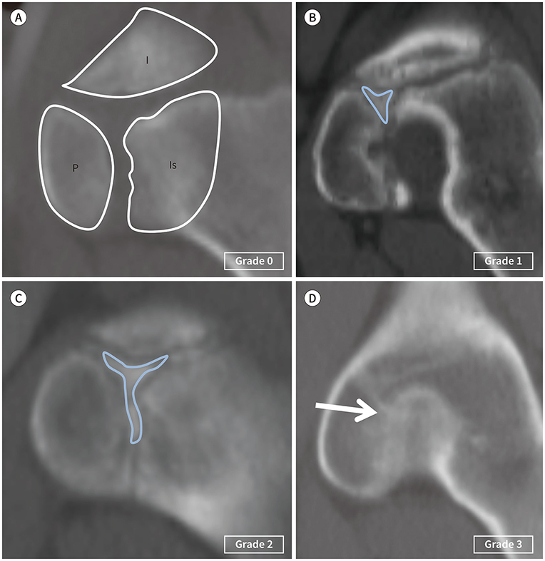
Fig. 1 TRC grading. Reformed sagittal, abdominal, pelvic CT shows the TRC ossification center (Grade 0–3). A. No ossification in the center of the three flanges of the TRC of the acetabulum. B. The beginning of ossification in the center of the three flanges (ossification center: annotated by blue colored triangle). C. The extension of the three flanges to the ileum, pubis and ischium (ossification center: annotated by the blue colored Y-shape). D. Maturation and complete fusion of the three flanges (arrow). I = ilium, Is = ischium, P = pubis, TRC= triradiate cartilage
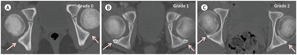
Fig. 2 PA grading. Axial abdominal, pelvic CT shows the PA ossification center (Grade 0–2). A. The entirely cartilaginous posterior wall and the invisible ossification center (arrows) surrounding the posterior column. B. The appearance of irregular ossification (arrows) in the posterior wall. C. The entire osseous posterior wall that has been fused with the posterior column (arrows). PA = posterior acetabular wall
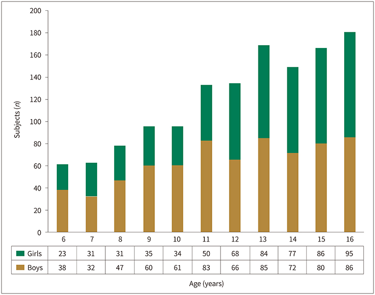
Fig. 3 Patient demographics (age and gender distribution of the number of CT scans). The total number of CT scans (n = 1324).
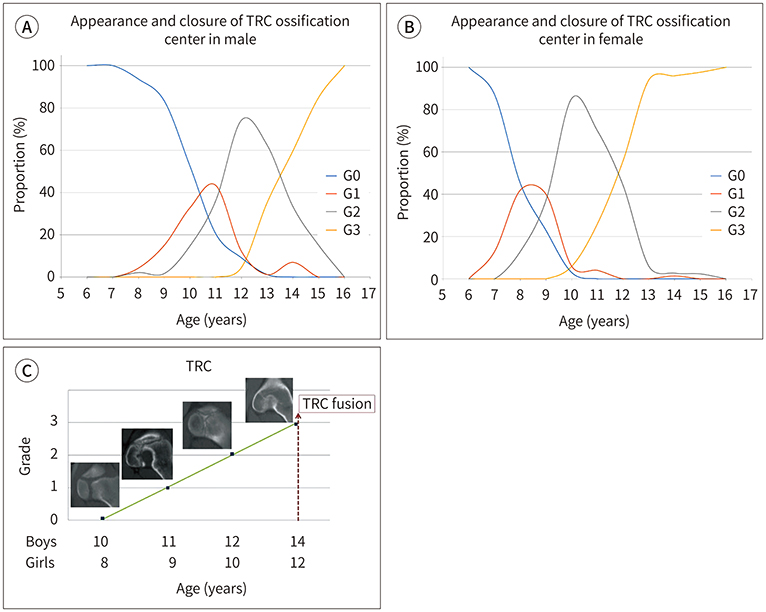
Fig. 4 Chronological age at appearance and closure of the TRC ossification center. The ossification centers of TRC of the boys first appear in the center of the three flanges (G1) at the age of 11 years and grow into three flanges (G2) at age 12, and complete bony fusion (G3) at age 14 (A, B). TRC maturation of the girls is exactly two years earlier than boys, revealing G1 at age 9; G2 at age 10; and G3, at age 12 (C). G0 = Grade 0, G1 = Grade 1, G2 = Grade 2, G3 = Grade 3, TRC= triradiate cartilage
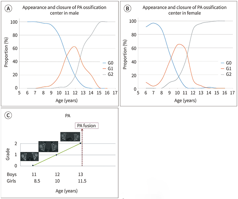
Fig. 5 Chronological age at appearance and closure of the PA ossification center. The PA of the boys starts to ossify at age 11 and completes fusion at age 13. The PA of the girls G1 is seen at age 10 and G2 at age 11.5 (A–C). G0 = Grade 0, G1 = Grade 1, G2 = Grade 2, PA = posterior acetabular wall
Reference
-
1. Canavese F, Charles YP, Dimeglio A. Skeletal age assessment from elbow radiographs. Review of the literature. Chir Organi Mov. 2008; 92:1–6.
Article2. Hacquebord JH, Leopold SS. In brief: the Risser classification: a classic tool for the clinician treating adolescent idiopathic scoliosis. Clin Orthop Relat Res. 2012; 470:2335–2338.
Article3. Charles YP, Diméglio A, Canavese F, Daures JP. Skeletal age assessment from the olecranon for idiopathic scoliosis at Risser grade 0. J Bone Joint Surg Am. 2007; 89:2737–2744.
Article4. Kim H, Kim HS, Moon ES, Yoon CS, Chung TS, Song HT, et al. Scoliosis imaging: what radiologists should know. Radiographics. 2010; 30:1823–1842.
Article5. Modi HN, Modi CH, Suh SW, Yang JH, Hong JY. Correlation and comparison of Risser sign versus bone age determination (TW3) between children with and without scoliosis in Korean population. J Orthop Surg Res. 2009; 4:36.
Article6. Satoh M. Bone age: assessment methods and clinical applications. Clin Pediatr Endocrinol. 2015; 24:143–152.
Article7. Paesano PL, Vigone MC, Siragusa V, Chiumello G, Del Maschio A, Mora S. Assessment of skeletal maturation in infants: comparison between two methods in hypothyroid patients. Pediatr Radiol. 1998; 28:622–626.
Article8. Girdany BR, Golden R. Centers of ossification of the skeleton. Am J Roentgenol Radium Ther Nucl Med. 1952; 68:922–924.9. Parvaresh KC, Upasani VV, Bomar JD, Pennock AT. Secondary Ossification Center appearance and closure in the pelvis and proximal femur. J Pediatr Orthop. 2018; 38:418–423.
Article10. Fabricant PD, Hirsch BP, Holmes I, Kelly BT, Lorich DG, Helfet DL, et al. A radiographic study of the ossification of the posterior wall of the acetabulum: implications for the diagnosis of pediatric and adolescent hip disorders. J Bone Joint Surg Am. 2013; 95:230–236.
Article11. Dimeglio A. Growth in pediatric orthopaedics. J Pediatr Orthop. 2001; 21:549–555.
Article12. Holden CP, Holman J, Herman MJ. Pediatric pelvic fractures. J Am Acad Orthop Surg. 2007; 15:172–177.
Article13. Liporace FA, Ong B, Mohaideen A, Ong A, Koval KJ. Development and injury of the triradiate cartilage with its effects on acetabular development: review of the literature. J Trauma. 2003; 54:1245–1249.14. Scheinfeld MH, Dym AA, Spektor M, Avery LL, Dym RJ, Amanatullah DF. Acetabular fractures: what radiologists should know and how 3D CT can aid classification. Radiographics. 2015; 35:555–577.
Article15. Bredella MA, Misra M, Miller KK, Madisch I, Sarwar A, Cheung A, et al. Distal radius in adolescent girls with anorexia nervosa: trabecular structure analysis with high-resolution flat-panel volume CT. Radiology. 2008; 249:938–946.
Article16. Lustrin ES, Karakas SP, Ortiz AO, Cinnamon J, Castillo M, Vaheesan K, et al. Pediatric cervical spine: normal anatomy, variants, and trauma. Radiographics. 2003; 23:539–560.
Article17. Bayaroğulları H, Yengil E, Davran R, Ağlagül E, Karazincir S, Balcı A. Evaluation of the postnatal development of the sternum and sternal variations using multidetector CT. Diagn Interv Radiol. 2014; 20:82–89.18. Breen MA, Tsai A, Stamm A, Kleinman PK. Bone age assessment practices in infants and older children among Society for Pediatric Radiology members. Pediatr Radiol. 2016; 46:1269–1274.
Article
- Full Text Links
-
- Actions
-
Cited
- CITED
-
- Close
- Share
-
- Similar articles
-
- Conservative Treatment of Physeal Bar after Triradiate Cartilage Injury
- Triradiate Approach in Surgical Treatment of Complex Fracture of Acetabulum
- The Role of Y and Greater Trochanteric Growth Cartilage upon the Acetabular Development of Rabbits: An Experimental Study
- Surgical Treatment of the Posterior wall Fracture of Acetabulum with Posterior Hip Dislocation
- A Case of Auricular Ossification
