J Korean Ophthalmol Soc.
2019 Jul;60(7):692-695. 10.3341/jkos.2019.60.7.692.
A Lacrimal Punctal Keratinizing Cyst of the Lower Lid
- Affiliations
-
- 1Department of Ophthalmology, Chungnam National University College of Medicine, Daejeon, Korea. sblee@cnu.ac.kr
- KMID: 2453021
- DOI: http://doi.org/10.3341/jkos.2019.60.7.692
Abstract
- PURPOSE
We report an unusual case of a keratinizing cyst on the lacrimal punctum.
CASE SUMMARY
A 49-year-old female presented with an outpouching punctal mass at the left lower lid that occurred a week prior to her visit. Histopathological examination revealed a cyst filled with keratin arranged in lamina and surrounding the bacterial colony. The epithelial wall was composed of multilaminar, keratinizing squamous epithelium without goblet cells. The features were consistent with a keratinizing cyst. There was no recurrence at 4 months after the excision, and the punctum was patent.
CONCLUSIONS
Keratinizing cyst should be considered as a differential diagnosis of the cystic mass of the punctum. Because it has an excellent prognosis after surgical resection, complete resection should be performed if a keratinizing cyst is suspected.
Keyword
MeSH Terms
Figure
-
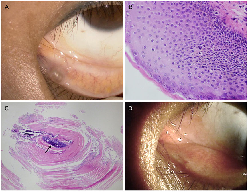
Figure 1 The keratinizing cyst of the left lower lid. (A) A photograph of the left lower eyelid showing a 2 mm, tense, whitish, non-vascular, smooth, cystic, dome-shaped mass at the region of lower punctum. (B) The epithelium was multilayered and showed a regular basaloid germinal layer without any goblet cells or any granular layer. Microphotograph showed keratin fibers on the stratified squamous epithelium (Haemotoxylin and Eosin [H&E] stain, ×400). (C) Microphotograph of the cystic content showing central bacterial colony (arrow) and surrounding keratin fibers arranged in laminar and wavy patterns (H&E stain, ×40). (D) There was no recurrence after 4 months.
Reference
-
1. Rumelt S, Pe'er J, Rubin PA. The clinicopathological spectrum of benign peripunctal tumours. Graefes Arch Clin Exp Ophthalmol. 2005; 243:113–119.
Article2. Ali MJ, Naik MN, Kaliki S. Punctal keratinizing cyst: a clinicopathological correlation of an exceptionally rare lacrimal disorder. Ophthalmic Plast Reconstr Surg. 2015; 31:e66–e68.3. Kamal S, Ali MJ, Naik MN. Punctal keratinizing cyst: report in a pediatric patient with fourier domain optical coherence tomography features. Ophthalmic Plast Reconstr Surg. 2015; 31:161–163.4. Yonekawa Y, Jakobiec FA, Zakka FR, Kim N. Keratinizing cyst of the lacrimal punctum. Cornea. 2013; 32:883–885.
Article5. Suh JY, Jung HM, Ahn HB, Kim MH. Clinical features and treatment of peripunctal tumors. J Korean Ophthalmol Soc. 2012; 53:918–923.
Article6. Wang L, Bi C, Wang T, et al. A coagulase-negative and non-haemolytic strain of Staphylococcus aureus for investigating the roles of SrtA in a murine model of bloodstream infection. Pathog Dis. 2015; 73:ftv042.7. Kuniyuki S, Yoshida Y, Maekawa N, Yamanaka K. Bacteriological study of epidermal cysts. Acta Derm Venereol. 2008; 88:23–25.
Article
- Full Text Links
-
- Actions
-
Cited
- CITED
-
- Close
- Share
-
- Similar articles
-
- Study on Dry Eye Syndrome and Lacrimal Punctal Size
- Study of lacrimal Punctal Size in Normal Adults
- Congenital Punctal Agenesis associated with Syndaotyly in a Family
- A Case of Lacrimal Gland Duct Cyst Associated with Ptosis
- Efficacy of Dacryoscintigraphy in Patients with Functional Block of Lacrimal Drainage System
