Analysis of Contractile Properties in Gastrocnemius, Tibialis Anterior Muscle of Amateur Male Soccer Players Using Tensiomyography
- Affiliations
-
- 1Department of Physical Education, Tongmyong University, Busan, Korea. hwangbg@tu.ac.kr
- KMID: 2452949
- DOI: http://doi.org/10.15384/kjhp.2019.19.2.114
Abstract
- BACKGROUND
Tensiomyography (TMG) is a relatively new technique that assesses the contractile properties of muscles in response to a single electrical stimulus. This study aimed to evaluate the contractile properties of the gastrocnemius and tibialis anterior (TA) muscles in amateur soccer players using TMG.
METHODS
We recruited 41 male soccer players (high school group, n=21; college group, n=20). The gastrocnemius medialis (GM), gastrocnemius lateralis (GL), and TA muscles of both lower extremities were assessed using TMG. The maximal displacement (Dm), delay time, contraction time (Tc), sustained time, and half-relaxation time were obtained and compared between the two groups.
RESULTS
First, both groups showed low Dm for the GM and TA muscles, which indicated high stiffness of the muscle tone. Second, the Tc and contraction velocity (Vc) were high for all muscles, except for the GL showing lower speed than the other muscles, which represented the sports-specific characteristics of the soccer players. Third, there were no significant differences in the measurement variables between the dominant and non-dominant sides, except for the Tc of the GM in high school athletes and Vc of the TA in college athletes.
CONCLUSIONS
These results reflected the sports-specific needs and characteristics of soccer players. A risk of injury is associated with a high degree of stiffness, and various methods for preventing it should be considered.
Keyword
MeSH Terms
Figure
-

Figure 1 (A) Components of tensiomyography system and (B) TMG record with parameters. Dm, maximal displacement; Tc, contraction time; Td, delay time; Tr, relaxation time; Ts, sustain time.
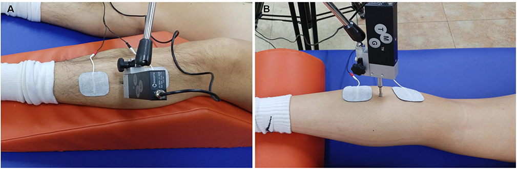
Figure 2 Positioning of the subject during tibialis anterior (A) and gastrocnemius medialis (B) measurements.
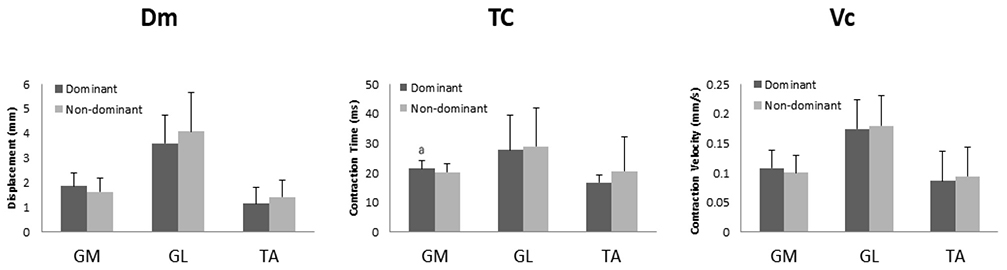
Figure 3 Comparison between dominant and non-dominant on Dm, Tc, Vc of high-school. Dm, displacement maximum; GL, gastrocnemius lateralis; GM, gastrocnemius medialis; TA, tibialis anterior; Tc, contraction time; Vc, contraction velocity. aP<0.05.

Figure 4 Comparison between dominant and non-dominant on Dm, Tc, Vc of collegiate. Dm, displacement maximum; GL, gastrocnemius lateralis; GM, gastrocnemius medialis; TA, tibialis anterior; Tc, contraction time; Vc, contraction velocity. aP<0.05.
Reference
-
1. Kim KJ. Effective training strategy for the improvement of exercise performance. J Coaching Dev. 2013; 15(1):72–83.2. Valenčič V, Knez N. Measuring of skeletal muscles' dynamic properties. Artif Organs. 1997; 21(3):240–242.
Article3. Tous-Fajardo J, Moras G, Rodríguez-Jiménez S, Usach R, Doutres DM, Maffiuletti NA. Inter-rater reliability of muscle contractile property measurements using non-invasive tensiomyography. J Electromyogr Kinesiol. 2010; 20(4):761–766.
Article4. Rusu LD, Cosma GG, Cernaianu SM, Marin MN, Rusu PF, Ciocănescu DP, et al. Tensiomyography method used for neuromuscular assessment of muscle training. J Neuroeng Rehabil. 2013; 10:67.
Article5. Križaj D, Šimunič B, Žagar T. Short-term repeatability of parameters extracted from radial displacement of muscle belly. J Electromyogr Kinesiol. 2008; 18(4):645–651.
Article6. Bangsbo J, Mohr M, Krustrup P. Physical and metabolic demands of training and match-play in the elite football player. J Sports Sci. 2006; 24(7):665–674.
Article7. Manolopoulos E, Papadopoulos C, Salonikidis K, Katartzi E, Poluha S. Strength training effects on physical conditioning and instep kick kinematics in young amateur soccer players during preseason. Percept Mot Skills. 2004; 99(2):701–710.
Article8. Kaminski TW, Buckley BD, Powers ME, Hubbard TJ, Ortiz C. Effect of strength and proprioception training on eversion to inversion strength ratios in subjects with unilateral functional ankle instability. Br J Sports Med. 2003; 37(5):410–415. discussion 415.9. Ebig M, Lephart SM, Burdett RG, Miller MC, Pincivero DM. The effect of sudden inversion stress on EMG activity of the peroneal and tibialis anterior muscles in the chronically unstable ankle. J Orthop Sports Phys Ther. 1997; 26(2):73–77.
Article10. Khamis S, Yizhar Z. Effect of feet hyperpronation on pelvic alignment in a standing position. Gait Posture. 2007; 25(1):127–134.
Article11. Chai JH, Kim BK, Kim C, Kim CH, Bae SW. Analysis of body-builder's skeletal muscle characteristics using tensiomyography. Korean J Sports Med. 2016; 34(2):146–152.
Article12. Eo ES, Hwang BG. The comparison of contractile properties between knee flexor and extnsor muscles in highschool basketball players using tensiomyography(TMG). J Sport Leis Stud. 2017; 69:387–394.13. Kim BK, Chai JH, Kim C, Kim CH, Bae SW. Analysis of lower extremity contraction according to gender using tensiomyography. Korean J Sports Med. 2017; 35(3):181–189.
Article14. Alentorn-Geli E, Alvarez-Diaz P, Ramon S, Marin M, Steinbacher G, Rius M, et al. Assessment of gastrocnemius tensiomyographic neuromuscular characteristics as risk factors for anterior cruciate ligament injury in male soccer players. Knee Surg Sports Traumatol Arthrosc. 2015; 23(9):2502–2507.
Article15. Carrasco L, Sañudo B, de Hoyo M, Pradas F, Da Silva ME. Effectiveness of low-frequency vibration recovery method on blood lactate removal, muscle contractile properties and on time to exhaustion during cycling at VO2 max power output. Eur J Appl Physiol. 2011; 111(9):2271–2279.16. Rey E, Lago-Peñas C, Lago-Ballesteros J. Tensiomyography of selected lower-limb muscles in professional soccer players. J Electromyogr Kinesiol. 2012; 22(6):866–872.
Article17. Rodríguez-Matoso D, Rodríguez-Ruiz D, Sarmiento S, Vaamonde D, Da Silva-Grigoletto ME, García-Manso JM. Reproducibility of muscle response measurements using tensiomyography in a range of positions. Rev Andal Med Deport. 2010; 3(3):81–86.18. Delagi EF, Lazzetti J, Perotto AO. Anatomical guide for the electromyographer: the limbs and trunk. 5th ed. Springfield: Charles C Thomas;2011. p. 5–85.19. Dahmane R, Valenčič V, Knez N, Eržen I. Evaluation of the ability to make non-invasive estimation of muscle contractile properties on the basis of the muscle belly response. Med Biol Eng Comput. 2001; 39(1):51–55.
Article20. Rodríguez-Ruiz D, García-Manso JM, Rodríguez-Matoso D, Sarmiento S, Da Silva-Grigoletto M, Pisot R. Effects of age and physical activity on response speed in knee flexor and extensor muscles. Eur Rev Aging Phys Act. 2013; 10(2):127.
Article21. Speed C. High-performance sports medicine. Clin Med (Lond). 2013; 13(1):47–49.
Article22. Reily T, Mujika I. Science and football in an applied context. ICSSPE Bull. 2006; 47:8–14.23. Pisot R, Narici MV, Simunic B, De Boer M, Seynnes O, Jurdana M, et al. Whole muscle contractile parameters and thickness loss during 35-day bed rest. Eur J Appl Physiol. 2008; 104(2):409–414.
Article24. Alvarez-Diaz P, Alentorn-Geli E, Ramon S, Marin M, Steinbacher G, Rius M, et al. Comparison of tensiomyographic neuromuscular characteristics between muscles of the dominant and non-dominant lower extremity in male soccer players. Knee Surg Sports Traumatol Arthrosc. 2016; 24(7):2259–2263.
Article25. Hunter AM, Galloway SD, Smith IJ, Tallent J, Ditroilo M, Fairweather MM, et al. Assessment of eccentric exercise-induced muscle damage of the elbow flexors by tensiomyography. J Electromyogr Kinesiol. 2012; 22(3):334–341.
Article26. Dahmane R, Djordjevič S, Šimunič B, Valenčič V. Spatial fiber type distribution in normal human muscle: histochemical and tensiomyographical evaluation. J Biomech. 2005; 38(12):2451–2459.27. Mitani Y. Gender-related differences in lower limb alignment, range of joint motion, and the incidence of sports injuries in Japanese university athletes. J Phys Ther Sci. 2017; 29(1):12–15.
Article
- Full Text Links
-
- Actions
-
Cited
- CITED
-
- Close
- Share
-
- Similar articles
-
- Electromyographic Analysis of Left Leg Muscle Activity during Golf Driver Swing
- The Effect of Tibial Lengthening on the Muscle: A comparison between the tibialis anterior and gastrocnemius muscle
- Effect of Elastic Compression Stocking and Kinesio Taping during Heel-raise Exercise on Muscle Activity, Mechanical Properties, and Muscle Fatigue in Healthy Women
- Analysis of Landing Error Scoring System during Drop Vertical Jump on Anterior Cruciate Ligament Injury Risk Factors in Female Ballet Dancers and Female Soccer Players
- Evaluation of Muscle Damage by Central Fatigue Using Tensiomyography
