Kosin Med J.
2019 Jun;34(1):78-82. 10.7180/kmj.2019.34.1.78.
A Case of Malignant Lymphoma Misdiagnosed as Acute Tonsillitis with Subsequent Lymphadenitis
- Affiliations
-
- 1Department of Otorhinolaryngology, Gyeongsang National University School of Medicine, Gyeongsang National University Hospital, Jinju, Korea. capetown@hanmail.net
- 2Department of Otorhinolaryngology, Eulji Medical Center, Eulji University School of Medicine, Seoul, Korea.
- 3Department of Otorhinolaryngology, Gyeongsang National University School of Medicine, Gyeongsang National University Changwon Hospital, Changwon, Korea.
- 4Institute of Health Sciences, College of Medicine, Gyeongsang National University, Jinju, Korea.
- KMID: 2451780
- DOI: http://doi.org/10.7180/kmj.2019.34.1.78
Abstract
- A 56-year-old female presented with clinical features of acute tonsillitis with subsequent cervical lymphadenitis. After taking empirical antibiotics for 1 week, the acute infection symptoms and signs were resolved. However, an asymmetric enlargement of the left palatine tonsil with ipsilateral neck swelling remained. Subsequent tonsillectomy and lymph node excisional biopsy were performed due to the possibility of malignancy. The patient was eventually diagnosed as malignant lymphoma according to pathological confirmation. We demonstrate the diagnostic challenges in such a rare case and emphasize the importance of differentiating malignant lymphoma from an atypically presenting acute infectious disease.
Keyword
MeSH Terms
Figure
-
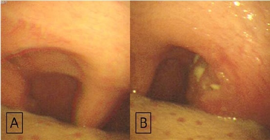
Fig. 1 Endoscopic examination of the tonsil. (A) Enlarged right palatine tonsil, (B) Asymmetrically enlarged left palatine tonsil with exudate on its surface.
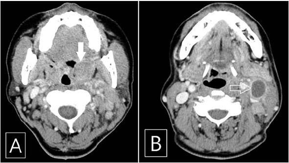
Fig. 2 Contrast enhanced computed tomography scan of the patient. (A) Hypertrophic and slightly enhanced left palatine tonsil (white arrow) and (B) Enlarged ipsilateral cervical lymph node which showed central low attenuated lesion (hollow arrow).
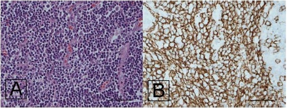
Fig. 3 Histopathological findings and immunohistochemical staining of the lymph node. (A) Diffusely infiltrated atypical lymphoid cells (H&E stain, ×400), (B) Strongly positive for CD20 immunohistochemical staining (×400).
Reference
-
1. Cooper JS, Porter K, Mallin K, Hoffman HT, Weber RS, Ang KK, et al. National Cancer Database report on cancer of the head and neck: 10-year update. Head Neck. 2009; 31:748–758.
Article2. Beasley MJ. Lymphoma of the thyroid and head and neck. Clin Oncol (R Coll Radiol). 2012; 24:345–351.
Article3. Laskar S, Mohindra P, Gupta S, Shet T, Muckaden MA. Non-Hodgkin lymphoma of the Waldeyer's ring: clinicopathologic and therapeutic issues. Leuk Lymphoma. 2008; 49:2263–2271.
Article4. Townsend W, Linch D. Hodgkin's lymphoma in adults. Lancet. 2012; 380:836–847.
Article5. Cinar F. Significance of asymptomatic tonsil asymmetry. Otolaryngol Head Neck Surg. 2004; 131:101–103.
Article6. Dolev Y, Daniel SJ. The presence of unilateral tonsillar enlargement in patients diagnosed with palatine tonsil lymphoma: experience at a tertiary care pediatric hospital. Int J Pediatr Otorhinolaryngol. 2008; 72:9–12.
Article7. Bartlett A, Bola S, Williams R. Acute tonsillitis and its complications: an overview. J R Nav Med Serv. 2015; 101:69–73.
Article8. Yellin SA, Weiss MH, Kraus DH, Papadopoulos EB. Tonsil lymphoma presenting as tonsillitis after bone marrow transplantation. Otolaryngol Head Neck Surg. 1995; 112:544–548.
Article9. Berkowitz RG, Mahadevan M. Unilateral tonsillar enlargement and tonsillar lymphoma in children. Ann Otol Rhinol Laryngol. 1999; 108:876–879.
Article10. Kallel S, Hadj Taieb H, Makni S, Ghorbel A. Lymphoma presenting as a peritonsillar abscess. Eur Ann Otorhinolaryngol Head Neck Dis. 2013; 130:337–339.
Article
- Full Text Links
-
- Actions
-
Cited
- CITED
-
- Close
- Share
-
- Similar articles
-
- Subacute Necrotizing Lymphadenitis in a 3 Year-Old Male Child
- A Case of Primary Malignant Lymphoma of the Orbit Treated by Radiotherapy
- Application of Gene Rearrangement Analysis for Diagnosis of Malignant Lymphoma
- A Case Report of Kikuchi-Fujimoto Disease with Immune Thrombocytopenic Purpura
- Histopathologic findings of necrotizing lymphadenitis
