Anat Biol Anthropol.
2019 Jun;32(2):69-75. 10.11637/aba.2019.32.2.69.
A Case Study of the Fetal Skeleton from a Joseon Period Cemetery
- Affiliations
-
- 1Department of History, College of Liberal Arts, Sejong University, Korea. redqin@sejong.ac.kr
- 2Department of Anthropology, College of Social Sciences, Seoul National University & ROK MND MAKRI Identification Center, Korea.
- KMID: 2451560
- DOI: http://doi.org/10.11637/aba.2019.32.2.69
Abstract
- In this study, the case of the burial of a couple with their perinatal child in a cemetery from the Joseon period (Eunpyeong site) of South Korea was examined. In archaeological populations, high mortality rates of young females are often associated with problems related to pregnancy and childbirth. However, discoveries of pregnant women and fetal skeletons are very rare in archaeological research. Here, we report the case of a burial of a pregnant female and her perinatal child from a Joseon period cemetery site. The gestational age of fetus was estimated to be between 8.5 months and 9.5 months based on the cranial size and long bone length. The pregnant female under study appears to have died with fetal remains in utero. An examination of this case did not provide evidence that stress of obstetrical event was the direct cause of death. The rare case presented here makes a valuable contribution to the literature on pregnancy and obstetrical issues in past populations.
Keyword
MeSH Terms
Figure
-
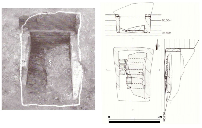
Fig. 1. Photo and drawing of burial 4-1-210, adapted from [9].
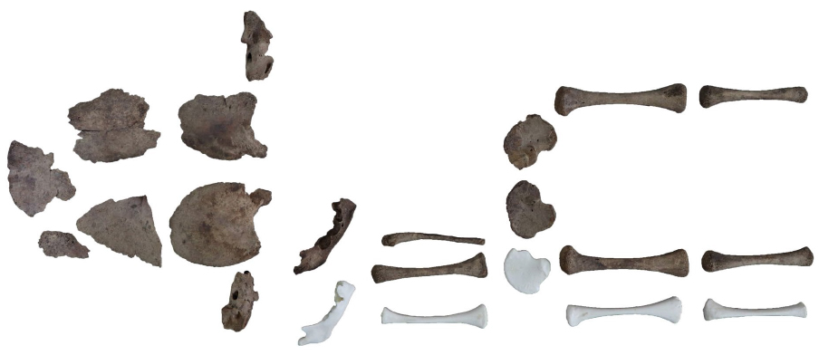
Fig. 2. Fetal skeleton from Burial 4-1-210 showing the degree of skeletal preservation (White skeleton: 8 months fetus from Bone Clones).
Reference
-
1. Tocheri MW, Dupras TL, Sheldrick P, Molto JE. Roman period fetal skeletons from the East Cemetery (Kellis 2) of Kellis, Egypt. Int J Osteoarchaeol. 2005; 15:326–341.
Article2. Ko I. Childbirth in archaeology. Journal of Humanities. 2014; 71:11–49. Korean.3. WHO Fact Sheet. Maternal mortality. World Health Organization; c2013 [cited 2019 June fifth]. Available from: http://www.who.int/mediacentre/factsheets/fs348/en/index.html.4. Arriaza B, Allison M, Gerszten E. Maternal mortality in pre-Columbian Indians of Arica, Chile. Am J Phys Anthropol. 1988; 77:35–41.
Article5. Cha M. Fertility, mortality and population growth in the late Joseon period: a study of life and death record in four genealogies from 1700 to 1899. Korean Demography. 2009; 32:113–137.6. The Samgang Institute of Cultural Properites. Nukdo Shell Mound V. 2006. Korean.7. Chang BS, UHM CS, Park CH, Kim HK, Jung HS, Ham JH, et al. Ultramicroscopic investigation of the preservation status of hair collected from a full-term, intrauterine baby mummy of the Joseon dynasty, Korea. Int J Osteoarchaeol. 2008; 18:624–631.
Article8. Woo EJ, Jeong YS, Cho GH, Pak S. Burial type and degenerative joint disease in the Joseon dynasty, Korea. J Korean Field Archaeol. 2011; 12:139–162.9. Central Institute of Cultural Heritage. Eunpyeong Jingwan dong Burial Site 4. 2009. p. 94. Korean.10. Pryse-Davies J, Smitham JH, Napier KA. Factors influencing development of secondary ossification centres in the fetus and newborn. Arch Dis Child. 1974; 49:425–431.11. O\'Rahilly R, Gardner E. The initial appearance of ossification in staged human embryos. Am J Anat. 1972; 134:291–308.
Article12. Mehta L, Singh HM. Determination of crown-rump length from fetal long bones: humerus and femur. Am J Phys Anthropol. 1972; 36:165–168.
Article13. Fazekas IG, Kósa F. In: Forensic fetal osteology. Budapest: Akadémiai Kiadó; 1978.14. Moorrees CFA, Fanning EA, Hunt EE. Formation and resorption of three deciduous teeth in children. Am J Phys Anthropol. 1963; 21:205–213.
Article15. Willis A, Oxenham MF. A case of maternal and perinatal death in Neolithic Southern Vietnam, c. 2100-1050 BCE. Int J Osteoarchaeol. 2013; 23:676–684.
Article16. Sherwood RJ, Meindl RS, Robinson HB, May RL. Fetal age: methods of estimation and effects of pathology. Am J Phys Anthropol. 2000; 113:305–315.
Article17. Scheuer JL, Musgrave JH, Evans SP. The estimation of late fetal and perinatal age from limb bone length by linear and logarithmic regression. Ann Hum Biol. 1980; 7:257–265.
Article18. Pak S, Woo EJ, Jeong Y. Report of physical anthropological analysis for human skeletal remains from Jingwan dong burial site, Eunpyeong, Seoul. In: Central Institute of Cultural Heritage. Eunpyeong Jingwan dong Burial Site 5. 2010. pp. 379-380. Korean.19. Brooks S, Suchey J. Skeletal age determination based on the os pubis: a comparison of the Acsádi-Nemeskéri and Suchey-Brooks methods. Hum Evol. 1990; 5:227–238.
Article20. Buikstra JE, Ubelaker DH. In: Standards for data collection from human skeletal remains. Arkansas Archeological Survey Research Series No. 44. Fayetteville: 1994.21. Meindl RS, Lovejoy CO. Ectocranial suture closure: a revised method for the determination of skeletal age at death based on the lateral-anterior sutures. Am J Phys Anthropol. 1985; 68:57–66.
Article22. Buckberry JL, Chamberlain AT. Age estimation from the auricular surface of the ilium: a revised method. Am J Phys Anthropol. 2002; 119:231–239.
Article23. Scott EC. Dental wear scoring technique. Am J Phys Anthropol. 1979; 51:213–217.
Article24. Owsley DW, Jantz RL. Long bone lengths and gestational age distributions of post-contact period Arikara Indian perinatal infant skeletons. Am J Phys Anthropol. 1985; 321–328.
Article25. Bello SM, Thomann A, Signoli M, Dutour O, Andrews P. Age and sex bias in the reconstruction of past population structures. Am J Phys Anthropol. 2006; 129:24–38.
Article26. Halcrow SE, Tayles N, Elliott GE. The bioarchaeology of fetuses. In: Han S, Betsinger TK, Scott AB, editors. The fetus: biology, culture, and society. Berghahn Books; 2017. pp. 83-111.27. Yi G. The lives and diseases of females during the latter half of the Joseon Dynasty as reconstructed with cases in Yeoksi Manpil. Korean J Med Hist. 2015; 24:497–532. Korean.28. Waldron I. Sex differences in human mortality: the role of genetic factors. Soc Sci Med. 1983; 17:321.
Article29. Cruz CB, Codinha S. Death of mother and child due to dystocia in 19th century Portugal. Int J Osteoarchaeol. 2010; 20:491–496.
Article30. Malgosa A, Alesan A, Safont S, Ballbé M, Ayala MM. A dystocic childbirth in the Spanish Bronze age. Int J Osteoarchaeol. 2004; 14:98–103.
Article
- Full Text Links
-
- Actions
-
Cited
- CITED
-
- Close
- Share
-
- Similar articles
-
- A Life and Death of the Leprosy Patients in Joseon Society Considered from the Anthropological Perspective
- Forensic Anthropological Study on Saw Marks Appearing on the Tibiae of a Joseon Skeleton
- Evidence of Periostitis in Joseon Dynasty Skeletons
- Stable isotope analysis of Joseon people skeletons from the cemeteries of Old Seoul City, the capital of Joseon Dynasty
- The surface and the back of late Joseon medicine - Centered on medical knowledge system -
