Role of Ultrasonography in Diagnosis and Treatment of Frozen Shoulder
- Affiliations
-
- 1Department of Rehabilitation Medicine, Daegu Catholic University School of Medicine, Daegu, Korea. parkgy@cu.ac.kr
- KMID: 2451506
- DOI: http://doi.org/10.4078/jrd.2019.26.3.149
Abstract
- Frozen shoulder (FS) is a common, painful and disabling condition of the shoulder. Patients usually present with an insidious onset of symptoms with gradual restriction and loss of shoulder mobility. FS can be categorized into primary and secondary types. The natural course of FS is characterized by the following 3 stages: the painful, the freezing/frozen, and the thawing phase based on the duration of symptoms, as well as pain and limitation of motion observed on physical examination. Diagnosis of FS is based on careful and accurate history taking and physical examination. Imaging modalities including arthrography, ultrasonography, and magnetic resonance imaging are useful in excluding concomitant painful conditions of the shoulder and in confirming FS. Ultrasonography is recommended as the first-line imaging modality to diagnose FS because it is noninvasive, it provides an easy comparison of ultrasonography parameters between the affected and unaffected sides, and it reflects the clinical characteristics of FS. The goal of treatment in patients with FS is pain reduction and restoration of normal function and mobility of the shoulder. Ultrasonography-guided glenohumeral joint injection, suprascapular nerve block, and distention arthrography achieve favorable therapeutic outcomes by virtue of greater accuracy. Ultrasonography and ultrasonography guided injections can accurately diagnose and effectively treat patients with FS.
Keyword
MeSH Terms
Figure
-

Figure 1 Ultrasonography measurement of coracohumeral ligament thickness. Longitudinal ultrasonography scan shows that the coracohumeral ligament thickness of the affected shoulder is thicker than that of the unaffected shoulder in the patient with frozen stage frozen shoulder.
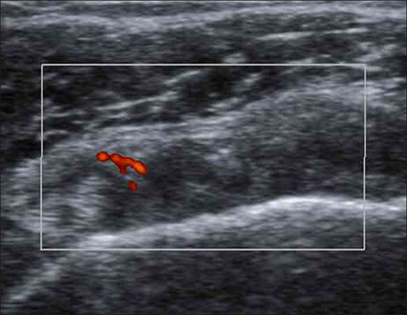
Figure 2 Transverse power Doppler ultrasonography scan shows hypervascularity in rotator cuff interval.
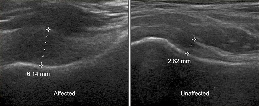
Figure 3 Ultrasonography measurement of axillary recess thickness. Longitudinal ultrasonography scan shows that the axillary recess thickness of the affected shoulder is thicker than that of the unaffected shoulder in the patient with frozen stage frozen shoulder. Axillary recess ratio is 2.34 (6.14/2.62 mm).
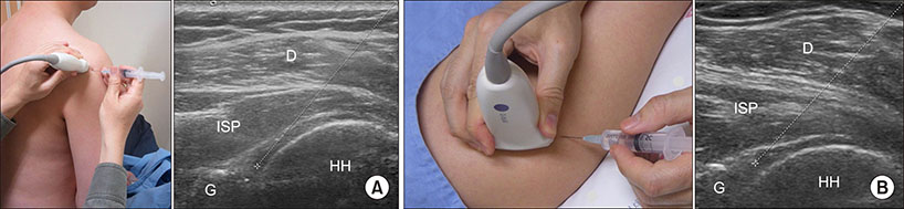
Figure 4 Ultrasonography guided glenohumeral joint injection using posterior approach in seated (A) and prone (B) position. D: deltoid, ISP: infraspinatus muscle, G: glenoid, HH: humeral head.
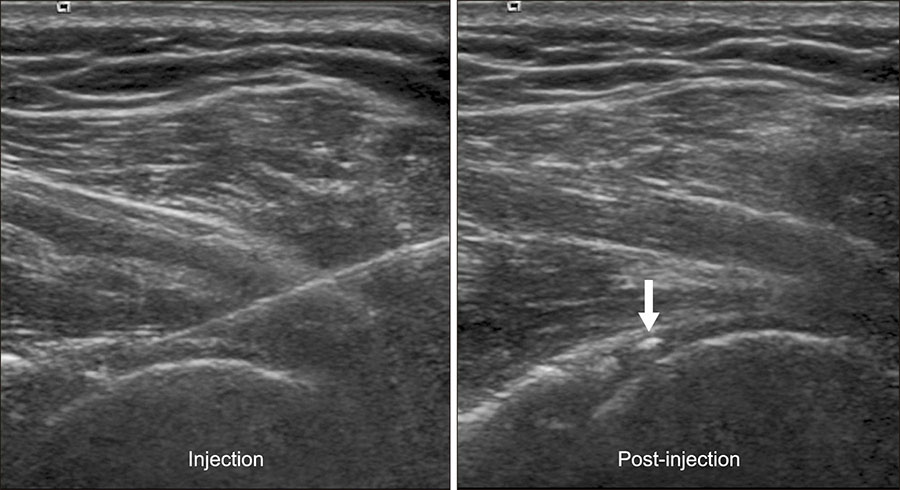
Figure 5 Ultrasonography guided glenohumeral joint injection using posterior approach in prone position. The needle tip is targeted at the point between the posterior labrum and posterior humeral head cartilage. The hyperechoic triamcinolone crystal (arrow) is detected in posterior glenohumeral joint after injection.

Figure 6 Distal (classic) suprascapular nerve block under ul- and serratus anterior muscle (SA). trasonography guidance. Needle is (arrow heads) approaching on the suprascapular nerve and artery (arrow) in suprascapular notch (open arrow head).

Figure 7 Proximal suprascapular nerve block under ultrasonography guidance, Needle tip (arrow) is located beneath proximal suprascapular nerve between omohyoid muscle (OM) and serratus anterior muscle (SA).
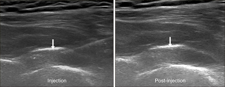
Figure 8 Glenohumeral distension arthrography under ultrasonography guidance. Capsular distension of posterior glenohumeral joint (arrow) is noted during and after distension arthrography.
Reference
-
1. Duplay S. [De la peri-arthrite scapulo-humerale et des raideurs de l’epaule qui en sont la consequence]. Arch Gen Med. 1872; 20:513–542.2. Codman EA. The shoulder: rupture of the supraspinatus tendon and other lesions in or about the subcromial bursa. Boston: Thomas Todd;1934. p. 216–224.3. Neviaser JS. Adhesive capsulitis of the shoulder. A study of the pathological findings in periarthritis of the shoulder. J Bone Joint Surg Am. 1945; 27:211–222.4. Lundberg BJ. The frozen shoulder. Clinical and radiographical observations. The effect of manipulation under general anesthesia. Structure and glycosaminoglycan content of the joint capsule Local bone metabolism. Acta Orthop Scand Suppl. 1969; 119:1–59.5. Zuckerman JD, Rokito A. Frozen shoulder: a consensus definition. J Shoulder Elbow Surg. 2011; 20:322–325.6. Reeves B. The natural history of the frozen shoulder syndrome. Scand J Rheumatol. 1975; 4:193–196.7. Hannafin JA, Chiaia TA. Adhesive capsulitis: a treatment approach. Clin Orthop Relat Res. 2000; (372):95–109.8. Cyriax JH. Textbook of orthopaedic medicine. Vol. 1, diagnosis of soft tissue lesions. 8th ed. London: Baillière Tindall;1982. p. 134–135.9. Loyd JA, Loyd HM. Adhesive capsulitis of the shoulder: arthrographic diagnosis and treatment. South Med J. 1983; 76:879–883.10. Neviaser TJ. Arthrography of the shoulder. Orthop Clin North Am. 1980; 11:205–217.11. Connell D, Padmanabhan R, Buchbinder R. Adhesive capsulitis: role of MR imaging in differential diagnosis. Eur Radiol. 2002; 12:2100–2106.12. Mengiardi B, Pfirrmann CW, Gerber C, Hodler J, Zanetti M. Frozen shoulder: MR arthrographic findings. Radiology. 2004; 233:486–492.13. Homsi C, Bordalo-Rodrigues M, da Silva JJ, Stump XM. Ultrasound in adhesive capsulitis of the shoulder: is assessment of the coracohumeral ligament a valuable diagnostic tool? Skeletal Radiol. 2006; 35:673–678.14. Kwon DR, Kim MY, Chae YJ, Park JS, Kim JS, Yi TI. Comparison of coracohumeral ligament thickness between asymptomatic shoulders and adhesive capsulitis in Korean. J Korean Acad Rehabil Med. 2009; 33:392–395.15. Lee JC, Sykes C, Saifuddin A, Connell D. Adhesive capsulitis: sonographic changes in the rotator cuff interval with arthroscopic correlation. Skeletal Radiol. 2005; 34:522–527.16. McNally EG, Rees JL. Imaging in shoulder disorders. Skeletal Radiol. 2007; 36:1013–1016.17. Walmsley S, Osmotherly PG, Walker CJ, Rivett DA. Power doppler ultrasonography in the early diagnosis of primary/idiopathic adhesive capsulitis: an exploratory study. J Manipulative Physiol Ther. 2013; 36:428–435.18. Park GY, Park JH, Kwon DR, Kwon DG, Park J. Do the findings of magnetic resonance imaging, arthrography, and ultrasonography reflect clinical impairment in patients with idiopathic adhesive capsulitis of the shoulder? Arch Phys Med Rehabil. 2017; 98:1995–2001.19. Park GY, Kwon DR. Application of real-time sonoelastography in musculoskeletal diseases related to physical medicine and rehabilitation. Am J Phys Med Rehabil. 2011; 90:875–886.20. Wu CH, Chen WS, Wang TG. Elasticity of the coracohumeral ligament in patients with adhesive capsulitis of the shoulder. Radiology. 2016; 278:458–464.21. Park GY, Lee JH, Kwon DG. Ultrasonographic measurement of the axillary recess thickness in an asymptomatic shoulder. Ultrasonography. 2017; 36:139–143.22. Park GY, Hwang SE. Comparison of intraarticular steroid injection with and without capsular distension in adhesive capsulitis of the shoulder. J Korean Acad Rehabil Med. 2000; 24:1174–1179.23. Oh JH, Oh CH, Choi JA, Kim SH, Kim JH, Yoon JP. Comparison of glenohumeral and subacromial steroid injection in primary frozen shoulder: a prospective, randomized short-term comparison study. J Shoulder Elbow Surg. 2011; 20:1034–1040.24. Dahan TH, Fortin L, Pelletier M, Petit M, Vadeboncoeur R, Suissa S. Double blind randomized clinical trial examining the efficacy of bupivacaine suprascapular nerve blocks in frozen shoulder. J Rheumatol. 2000; 27:1464–1469.25. Ko KP, Kang DH, Shin BK. The proximal approach in an ultrasound-guided suprascapular nerve block. J Korean Orthop Assoc. 2017; 52:521–528.26. Rothe C, Steen-Hansen C, Lund J, Jenstrup MT, Lange KH. Ultrasound-guided block of the suprascapular nerve - a volunteer study of a new proximal approach. Acta Anaesthesiol Scand. 2014; 58:1228–1232.27. Buchbinder R, Green S, Forbes A, Hall S, Lawler G. Arthrographic joint distension with saline and steroid improves function and reduces pain in patients with painful stiff shoulder: results of a randomised, double blind, placebo controlled trial. Ann Rheum Dis. 2004; 63:302–309.28. Park GY, Kwon DR, Kwon DG, Rim JH. Comparison of therapeutic effectiveness between shoulder distention arthrography with translation mobilization and distention arthrography alone in patients with frozen shoulder. Ann Rehabil Med. 2018; 42:76–84.29. Ekelund AL, Rydell N. Combination treatment for adhesive capsulitis of the shoulder. Clin Orthop Relat Res. 1992; 105–109.30. Mitra R, Harris A, Umphrey C, Smuck M, Fredericson M. Adhesive capsulitis: a new management protocol to improve passive range of motion. PM R. 2009; 1:1064–1068.
- Full Text Links
-
- Actions
-
Cited
- CITED
-
- Close
- Share
-
- Similar articles
-
- Changes of rotator Cuff using Ultrasonography in Frozen Shoulder
- The therapeutic effect of interventional microadhesiolysis and nerve stimulation (IMNS) under ultrasonographic guide in frozen shoulder patient: A case report
- Ultrasound Guidance for Intra-Articular Shoulder Injections for Frozen Shoulder
- Comparison of Suprascapular Nerve Block and Shoulder Joint Injection for Treatment of Frozen Shoulder
- Ultrasonography for Diagnosing Sports-Related Shoulder Pain
