Korean J Radiol.
2016 Jun;17(3):330-338. 10.3348/kjr.2016.17.3.330.
Comparison of Iohexol-380 and Iohexol-350 for Coronary CT Angiography: A Multicenter, Randomized, Double-Blind Phase 3 Trial
- Affiliations
-
- 1Department of Radiology, Seoul National University Hospital, Seoul 03080, Korea. whal.lee@gmail.com
- 2Department of Radiology, Ajou University School of Medicine, Suwon 16499, Korea.
- 3Department of Radiology, Chungbuk National University Hospital, Cheongju 28644, Korea.
- 4Department of Radiology, Yonsei University Wonju College of Medicine, Wonju Severance Christian Hospital, Wonju 26426, Korea.
- 5Department of Radiology, Ewha Womans University Mokdong Hospital, Seoul 07985, Korea.
- 6Department of Radiology, Gachon University Gil Medical Center, Incheon 21565, Korea.
- 7Department of Radiology, Hanyang University Seoul Hospital, Seoul 04763, Korea.
- 8Department of Radiology, Korea University Anam Hospital, Seoul 02841, Korea.
- 9Department of Radiology, Korea University Guro Hospital, Seoul 08308, Korea.
- 10Department of Radiology, Soonchunhyang University Bucheon Hospital, Bucheon 14584, Korea.
- 11Department of Radiology, Hallym University Sacred Heart Hospital, Anyang 14068, Korea.
- 12Department of Radiology, Chonbuk National University Medical School and Hospital, Institute of Medical Science, Research Institute of Clinical Medicine and Biomedical Research Institute, Jeonju 54907, Korea.
- 13Department of Radiology, Severance Hospital, Yonsei University Health System, Seoul 03722, Korea.
- 14Department of Radiology, Seoul National University Bundang Hospital, Seongnam 13620, Korea.
- KMID: 2451408
- DOI: http://doi.org/10.3348/kjr.2016.17.3.330
Abstract
OBJECTIVE
This multi-center, randomized, double-blind, phase 3 trial was conducted to compare the safety and efficacy of contrast agents iohexol-380 and iohexol-350 for coronary CT angiography in healthy subjects.
MATERIALS AND METHODS
Volunteers were randomized to receive 420 mgI/kg of either iohexol-350 or iohexol-380 using a flow rate of 4 mL/sec. All adverse events were recorded. Two blinded readers independently reviewed the CT images and conflicting results were resolved by a third reader. Luminal attenuations (ascending aorta, left main coronary artery, and left ventricle) in Hounsfield units (HUs) and image quality on a 4-point scale were calculated.
RESULTS
A total of 225 subjects were given contrast media (115 with iohexol-380 and 110 with iohexol-350). There was no difference in number of adverse drug reactions between groups: 75 events in 56 (48.7%) of 115 subjects in the iohexol-380 group vs. 74 events in 51 (46.4%) of 110 subjects in the iohexol-350 group (p = 0.690). No severe adverse drug reactions were recorded. Neither group showed an increase in serum creatinine. Significant differences in mean density between the groups was found in the ascending aorta: 375.8 ± 71.4 HU with iohexol-380 vs. 356.3 ± 61.5 HU with iohexol-350 (p = 0.030). No significant differences in image quality scores between both groups were observed for all three anatomic evaluations (all, p > 0.05).
CONCLUSION
Iohexol-380 provides improved enhancement of the ascending aorta and similar attenuation of the coronary arteries without any increase in adverse drug reactions, as compared with iohexol-350 using an identical amount of total iodine.
Keyword
MeSH Terms
-
Adult
Aged
Aorta/diagnostic imaging
Computed Tomography Angiography/*methods
Contrast Media/adverse effects/*chemistry
Coronary Vessels/*diagnostic imaging
Creatinine/blood
Double-Blind Method
Electrocardiography
Female
Gastrointestinal Diseases/etiology
Healthy Volunteers
Humans
Image Interpretation, Computer-Assisted
Iohexol/adverse effects/*chemistry
Male
Middle Aged
Nervous System Diseases/etiology
Skin Diseases/etiology
Young Adult
Contrast Media
Creatinine
Iohexol
Figure
-
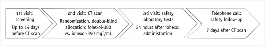
Fig. 1 Study flow.
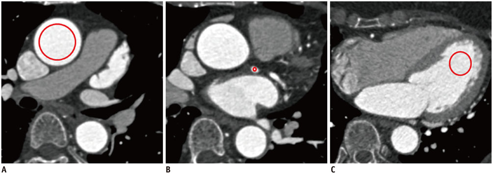
Fig. 2 Assessment of attenuation in three anatomical regions including ascending aorta (A), left main coronary artery (B), and left ventricular cavity (C). Circles indicate regions of interest placed at each region.
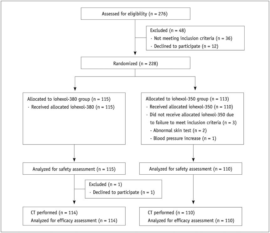
Fig. 3 Flow diagram of study patients.
Cited by 1 articles
-
Incidence of Breakthrough Reaction in Patients with Prior Acute Allergic-Like Reactions to Iodinated Contrast Media according to the Administration Route
Yeon Soo Kim, Young Hun Choi, Yeon Jin Cho, Seunghyun Lee, Soon Ho Yoon, Chang Min Park, Hye Ryun Kang
Korean J Radiol. 2018;19(2):352-357. doi: 10.3348/kjr.2018.19.2.352.
Reference
-
1. Kim YJ, Yong HS, Kim SM, Kim JA, Yang DH, Hong YJ. Korean Society of Radiology. Korean Society of Cardiology. Korean guidelines for the appropriate use of cardiac CT. Korean J Radiol. 2015; 16:251–285.2. Yoon YE, Lim TH. Current roles and future applications of cardiac CT: risk stratification of coronary artery disease. Korean J Radiol. 2014; 15:4–11.3. Pugliese F. Which contrast agent for coronary ct angiography?. RAD Magazine. 2008. 10.4. Cademartiri F, Mollet NR, Lemos PA, Saia F, Midiri M, de Feyter PJ, et al. Higher intracoronary attenuation improves diagnostic accuracy in MDCT coronary angiography. AJR Am J Roentgenol. 2006; 187:W430–W433.5. Becker CR, Hong C, Knez A, Leber A, Bruening R, Schoepf UJ, et al. Optimal contrast application for cardiac 4-detector-row computed tomography. Invest Radiol. 2003; 38:690–694.6. Fleischmann D. Use of high concentration contrast media: principles and rationale-vascular district. Eur J Radiol. 2003; 45:Suppl 1. S88–S93.7. Herman S. Computed tomography contrast enhancement principles and the use of high-concentration contrast media. J Comput Assist Tomogr. 2004; 28:Suppl 1. S7–S11.8. Bae KT. Peak contrast enhancement in CT and MR angiography: when does it occur and why? Pharmacokinetic study in a porcine model. Radiology. 2003; 227:809–881.9. Cademartiri F, de Monye C, Pugliese F, Mollet NR, Runza G, van der Lugt A, et al. High iodine concentration contrast material for noninvasive multislice computed tomography coronary angiography: iopromide 370 versus iomeprol 400. Invest Radiol. 2006; 41:349–353.10. Kim EY, Yeh DW, Choe YH, Lee WJ, Lim HK. Image quality and attenuation values of multidetector CT coronary angiography using high iodine-concentration contrast material: a comparison of the use of iopromide 370 and iomeprol 400. Acta Radiol. 2010; 51:982–989.11. Lim J, Park EA, Lee W, Shim H, Chung JW. Image quality and radiation reduction of 320-row area detector CT coronary angiography with optimal tube voltage selection and an automatic exposure control system: comparison with body mass index-adapted protocol. Int J Cardiovasc Imaging. 2015; 31:Suppl 1. 23–30.12. Tsai IC, Lee T, Tsai WL, Chen MC, Wu MJ, Lee WL, et al. Contrast enhancement in cardiac MDCT: comparison of iodixanol 320 versus iohexol 350. AJR Am J Roentgenol. 2008; 190:W47–W53.13. Cademartiri F, Mollet NR, van der Lugt A, McFadden EP, Stijnen T, de Feyter PJ, et al. Intravenous contrast material administration at helical 16-detector row CT coronary angiography: effect of iodine concentration on vascular attenuation. Radiology. 2005; 236:661–665.14. Brink JA. Use of high concentration contrast media (HCCM): principles and rationale--body CT. Eur J Radiol. 2003; 45:Suppl 1. S53–S58.15. Rist C, Nikolaou K, Kirchin MA, van Gessel R, Bae KT, von Ziegler F, et al. Contrast bolus optimization for cardiac 16-slice computed tomography: comparison of contrast medium formulations containing 300 and 400 milligrams of iodine per milliliter. Invest Radiol. 2006; 41:460–467.16. Bae KT, Heiken JP. Scan and contrast administration principles of MDCT. Eur Radiol. 2005; 15:Suppl 5. E46–E59.17. Cademartiri F, van der Lugt A, Luccichenti G, Pavone P, Krestin GP. Parameters affecting bolus geometry in CTA: a review. J Comput Assist Tomogr. 2002; 26:598–607.18. Behrendt FF, Pietsch H, Jost G, Sieber MA, Keil S, Plumhans C, et al. Intra-individual comparison of different contrast media concentrations (300mg, 370mg and 400mg iodine) in MDCT. Eur Radiol. 2010; 20:1644–1650.19. Faggioni L, Neri E, Sbragia P, Pascale R, D'Errico L, Caramella D, et al. 80-kV pulmonary CT angiography with 40mL of iodinated contrast material in lean patients: comparison of vascular enhancement with iodixanol (320mg I/mL)and iomeprol (400mg I/mL). AJR Am J Roentgenol. 2012; 199:1220–1225.20. Han JK, Kim AY, Lee KY, Seo JB, Kim TK, Choi BI, et al. Factors influencing vascular and hepatic enhancement at CT: experimental study on injection protocol using a canine model. J Comput Assist Tomogr. 2000; 24:400–406.
- Full Text Links
-
- Actions
-
Cited
- CITED
-
- Close
- Share
-
- Similar articles
-
- Encephalopathy with Status Epilepticus after Iohexol Cervical Myelography
- Generalized Tonic-Clonic Seizure following Myelography with Iohexol (Omnipaque.): A Case Report
- Acute Post-Myelographic Meningitis with Iohexol
- A case of chemical meningitis after myelography
- Three-dimensional CT angiography of the canine hepatic vasculature
