The use of human mature cystic ovarian teratoma as a model to study the development of human lymphatics
- Affiliations
-
- 1Department of Anatomy, Jordan University of Science and Technology, Irbid, Jordan.
- 2Department of Obstetrics and Gynecology, Jordan University of Science and Technology, Irbid, Jordan. zoamarin@hotmail.com
- KMID: 2451235
- DOI: http://doi.org/10.5115/acb.2017.50.2.104
Abstract
- The angiogenic theory to the development of human lymphatics is not clear. The objective of this study was to investigate the development of human lymphatics. Semi-thin and thin paraffin sections from human mature cystic ovarian teratoma tissues were studied using light and electron microscopy. Lymphatics were formed by the differentiation of mesenchymal cells that gradually acquired morphological features of endothelial cells. It is suggested that in human mature cystic ovarian teratoma the lymphatic endothelium develops from mesenchymal cells, and not from cells derived from mature endothelium of a preexisting vein or lymphatic.
Keyword
MeSH Terms
Figure
-
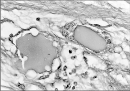
Fig. 1 Light micrograph showing two vessel-like structures, with a center of amorphous eosinophilic substance surrounded by mesenchymal cells lining the center and expanding slender cell processes in the vicinity (×400).
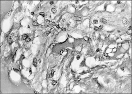
Fig. 2 Light micrograph showing a center of amorphous eosinophilic substance lined by several cells. Some cells have a thin profile creating a single opening while other cells have rounded profiles (×400).

Fig. 3 A semi-then section showing a lymphatic vessel lined by endothelial like cells surrounded by openings of variable size. The endothelial lining on the lower right side appears more differentiated than the other part (×400).
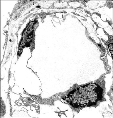
Fig. 4 Electron micrograph showing a well-developed lymphatic vessel with flattened endothelial cells forming a continuous layer (×100,000).
Reference
-
1. Fajardo LF. The complexity of endothelial cells: a review. Am J Clin Pathol. 1989; 92:241–250.2. Leak LV. The structure of lymphatic capillaries in lymph formation. Fed Proc. 1976; 35:1863–1871.3. Canzonieri V, Volpe R, Gloghini A, Carbone A. Sieve-like areas in mature cystic teratomas of the ovary: a histochemical and immunohistochemical study of 7 cases. Pathologica. 1994; 86:43–46.4. Klika E, Antaliková L, Rychter Z, Jelínek R. Inception and manner of development of the lymph vessels in the chick embryo heart. Lymphology. 1972; 5:137–148.5. Al-Jomard RM, Scothorne RJ. The development of lymphatics in the rat liver. In : Proceedings of the Anatomical Society of Great Britain and Ireland; 1986 June; Leicester, UK. London: Taylor and Francis;1986. p. 40.
- Full Text Links
-
- Actions
-
Cited
- CITED
-
- Close
- Share
-
- Similar articles
-
- A Case of Advanced Squamous Cell Carcinoma Arising in Mature Cystic Teratoma of the Ovary
- A Case of Adenocarcinoma Arising in Mature Cystic Teratoma of the Ovary
- Traumatic Rupture of a Mature Cystic Teratoma of the Ovary
- A Case of Squamous Cell Carcinoma Arising in Mature Cystic Teratoma of the Ovary
- Squamous Cell Carcinoma, Adenocarcinoma and Papillary Cell Carcinoma Arising from the Mature Cystic Teratoma of the Ovary: Case Report
