Imaging Sci Dent.
2019 Jun;49(2):115-122. 10.5624/isd.2019.49.2.115.
Comparison of cone-beam computed tomography and panoramic radiography in the evaluation of maxillary sinus pathology related to maxillary posterior teeth: Do apical lesions increase the risk of maxillary sinus pathology?
- Affiliations
-
- 1Department of Endodontics, Faculty of Dentistry, Necmettin Erbakan University, Konya, Turkey.
- 2Department of Oral and Maxillofacial Radiology, Faculty of Dentistry, Necmettin Erbakan University, Konya, Turkey. dishekmelek@gmail.com
- KMID: 2450177
- DOI: http://doi.org/10.5624/isd.2019.49.2.115
Abstract
- PURPOSE
The aims of this study were first, to compare panoramic radiography with cone-beam computed tomography (CBCT) for evaluating topographic relationships, such as the classification of maxillary posterior teeth and their distance to the maxillary sinus floor; and second, to determine the relationship between maxillary sinus pathology and the presence of apical lesions.
MATERIALS AND METHODS
In total, 285 paired CBCT and panoramic radiography records of patients (570 maxillary sinuses) were retrospectively analyzed. Both imaging modalities were used to determine the topographic relationship of the maxillary posterior teeth to the sinus floor. Mucosal thickening >2 mm was considered a pathological state. Data were analyzed using the chi-square, Wilcoxon, and Mann-Whitney U tests. Odds ratios (ORs) and confidence intervals (CIs) were calculated.
RESULTS
The closest vertical distance measurements made between posterior maxillary teeth roots and the maxillary sinus on panoramic radiography and CBCT scans showed statistically significant differences from each other (P<0.05). Compared to panoramic radiography, CBCT showed higher mean values for the distance between the maxillary sinus floor and maxillary posterior teeth roots. The CBCT images showed that at least 1 apical lesion adjacent to the right maxillary sinus increased the risk of maxillary sinus pathology by 2.37 times (OR, 2.37; 95% CI, 1.58-3.55, P<0.05).
CONCLUSION
Panoramic radiography might lead to unreliable diagnoses when evaluating the distance between the sinus floor and posterior roots of the maxillary teeth. Periapical lesions anatomically associated with maxillary sinuses were a risk factor for sinus mucosal thickening.
MeSH Terms
Figure
-
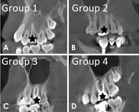
Fig. 1 Grouping of teeth on sagittal cone-beam computed tomography slices by apical condition. A, B, C, and D represent groups 1, 2, 3, and 4, respectively.

Fig. 2 Measurement of maxillary sinus mucosal thickening on a sagittal cone-beam computed tomography slice.
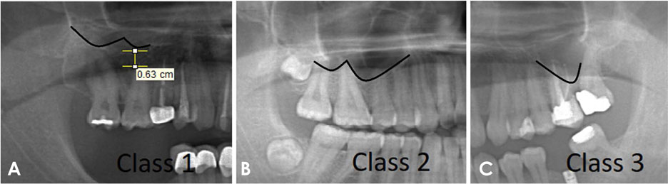
Fig. 3 Classification of the topographic relationships of maxillary first molars with the maxillary sinus on panoramic radiographs. A, B, and C represent classes 1, 2, and 3, respectively.

Fig. 4 Classification of the topographic relationships of maxillary first molars with the maxillary sinus on sagittal cone-beam computed tomography slices. A, B, and C represent classes 1, 2, and 3, respectively. CBCT, cone-beam computed tomography.
Reference
-
1. Lu Y, Liu Z, Zhang L, Zhou X, Zheng Q, Duan X, et al. Associations between maxillary sinus mucosal thickening and apical periodontitis using cone-beam computed tomography scanning: a retrospective study. J Endod. 2012; 38:1069–1074.
Article2. Melén I, Lindahl L, Andréasson L, Rundcrantz H. Chronic maxillary sinusitis. Definition, diagnosis and relation to dental infections and nasal polyposis. Acta Otolaryngol. 1986; 101:320–327.3. Abrahams JJ, Glassberg RM. Dental disease: a frequently unrecognized cause of maxillary sinus abnormalities? AJR Am J Roentgenol. 1996; 166:1219–1223.
Article4. Doud Galli SK, Lebowitz RA, Giacchi RJ, Glickman R, Jacobs JB. Chronic sinusitis complicating sinus lift surgery. Am J Rhinol. 2001; 15:181–186.
Article5. Kretzschmar DP, Kretzschmar JL. Rhinosinusitis: review from a dental perspective. Oral Surg Oral Med Oral Pathol Oral Radiol Endod. 2003; 96:128–135.
Article6. Savolainen S, Eskelin M, Jousimies-Somer H, Ylikoski J. Radiological findings in the maxillary sinuses of symptomless young men. Acta Otolaryngol Suppl. 1997; 529:153–157.
Article7. Vallo J, Suominen-Taipale L, Huumonen S, Soikkonen K, Norblad A. Prevalence of mucosal abnormalities of the maxillary sinus and their relationship to dental disease in panoramic radiography: results from the Health 2000 Health Examination Survey. Oral Surg Oral Med Oral Pathol Oral Radiol Endod. 2010; 109:e80–e87.
Article8. Ren S, Zhao H, Liu J, Wang Q, Pan Y. Significance of maxillary sinus mucosal thickening in patients with periodontal disease. Int Dent J. 2015; 65:303–310.
Article9. Shahbazian M, Vandewoude C, Wyatt J, Jacobs R. Comparative assessment of panoramic radiography and CBCT imaging for radiodiagnostics in the posterior maxilla. Clin Oral Investig. 2014; 18:293–300.
Article10. von Arx T, Fodich I, Bornstein MM. Proximity of premolar roots to maxillary sinus: a radiographic survey using cone-beam computed tomography. J Endod. 2014; 40:1541–1548.11. Sharan A, Madjar D. Correlation between maxillary sinus floor topography and related root position of posterior teeth using panoramic and cross-sectional computed tomography imaging. Oral Surg Oral Med Oral Pathol Oral Radiol Endod. 2006; 102:375–381.
Article12. Mozzo P, Procacci C, Tacconi A, Martini PT, Andreis IA. A new volumetric CT machine for dental imaging based on the cone-beam technique: preliminary results. Eur Radiol. 1998; 8:1558–1564.
Article13. Bremke M, Sesterhenn AM, Murthum T, Al Hail A, Bien S, Werner JA. Digital volume tomography (DVT) as a diagnostic modality of the anterior skull base. Acta Otolaryngol. 2009; 129:1106–1114.
Article14. Maestre-Ferrín L, Galán-Gil S, Carrillo-García C, Peñarrocha-Diago M. Radiographic findings in the maxillary sinus: comparison of panoramic radiography with computed tomography. Int J Oral Maxillofac Implants. 2011; 26:341–346.15. Shanbhag S, Karnik P, Shirke P, Shanbhag V. Association between periapical lesions and maxillary sinus mucosal thickening: a retrospective cone-beam computed tomographic study. J Endod. 2013; 39:853–857.
Article16. Ludlow JB, Ivanovic M. Comparative dosimetry of dental CBCT devices and 64-slice CT for oral and maxillofacial radiology. Oral Surg Oral Med Oral Pathol Oral Radiol Endod. 2008; 106:106–114.
Article17. Howe RB. First molar radicular bone near the maxillary sinus: a comparison of CBCT analysis and gross anatomic dissection for small bony measurement. Oral Surg Oral Med Oral Pathol Oral Radiol Endod. 2009; 108:264–269.
Article18. Hauman CH, Chandler NP, Tong DC. Endodontic implications of the maxillary sinus: a review. Int Endod J. 2002; 35:127–141.
Article19. Lofthag-Hansen S, Huumonen S, Gröndahl K, Gröndahl HG. Limited cone-beam CT and intraoral radiography for the diagnosis of periapical pathology. Oral Surg Oral Med Oral Pathol Oral Radiol Endod. 2007; 103:114–119.
Article20. Oberli K, Bornstein MM, von Arx T. Periapical surgery and the maxillary sinus: radiographic parameters for clinical outcome. Oral Surg Oral Med Oral Pathol Oral Radiol Endod. 2007; 103:848–853.
Article21. Eberhardt JA, Torabinejad M, Christiansen EL. A computed tomographic study of the distances between the maxillary sinus floor and the apices of the maxillary posterior teeth. Oral Surg Oral Med Oral Pathol. 1992; 73:345–346.
Article22. Kwak HH, Park HD, Yoon HR, Kang MK, Koh KS, Kim HJ. Topographic anatomy of the inferior wall of the maxillary sinus in Koreans. Int J Oral Maxillofac Surg. 2004; 33:382–388.
Article23. Nishikawa K, Suehiro A, Sekine H, Kousuge Y, Wakoh M, Sano T. Is linear distance measured by panoramic radiography reliable? Oral Radiol. 2010; 26:16–19.
Article24. Lopes LJ, Gamba TO, Bertinato JV, Freitas DQ. Comparison of panoramic radiography and CBCT to identify maxillary posterior roots invading the maxillary sinus. Dentomaxillofac Radiol. 2016; 45:20160043.
Article25. Brüllmann DD, Schmidtmann I, Hornstein S, Schulze RK. Correlation of cone beam computed tomography (CBCT) findings in the maxillary sinus with dental diagnoses: a retrospective cross-sectional study. Clin Oral Investig. 2012; 16:1023–1029.
Article26. Pommer B, Unger E, Sütö D, Hack N, Watzek G. Mechanical properties of the Schneiderian membrane in vitro. Clin Oral Implants Res. 2009; 20:633–637.27. Aimetti M, Massei G, Morra M, Cardesi E, Romano F. Correlation between gingival phenotype and Schneiderian membrane thickness. Int J Oral Maxillofac Implants. 2008; 23:1128–1132.28. Cagici CA, Yilmazer C, Hurcan C, Ozer C, Ozer F. Appropriate interslice gap for screening coronal paranasal sinus tomography for mucosal thickening. Eur Arch Otorhinolaryngol. 2009; 266:519–525.
Article29. Brüllmann D, Schulze RK. Spatial resolution in CBCT machines for dental/maxillofacial applications - what do we know today? Dentomaxillofac Radiol. 2015; 44:20140204.30. Temmerman A, Hertelé S, Teughels W, Dekeyser C, Jacobs R, Quirynen M. Are panoramic images reliable in planning sinus augmentation procedures? Clin Oral Implants Res. 2011; 22:189–194.
Article
- Full Text Links
-
- Actions
-
Cited
- CITED
-
- Close
- Share
-
- Similar articles
-
- Comparison of panoramic radiography and cone beam computed tomography for assessing the relationship between the maxillary sinus floor and maxillary molars
- Comparison of panoramic radiography and cone-beam computed tomography for assessing radiographic signs indicating root protrusion into the maxillary sinus
- The value of panoramic radiography in assessing maxillary sinus inflammation
- Assessment of maxillary third molars with panoramic radiography and cone-beam computed tomography
- Maxillary sinus septa: comparison between panoramic radiography and CBCT
