Imaging Sci Dent.
2019 Jun;49(2):97-102. 10.5624/isd.2019.49.2.97.
The influence of different scan resolutions on the detection of proximal caries lesions
- Affiliations
-
- 1Division of Oral Radiology, Department of Oral Diagnosis, Piracicaba Dental School, University of Campinas, Piracicaba, São Paulo, Brazil. haiter@fop.unicamp.br
- 2Section of Oral Radiology, Department of Dentistry and Oral Health, University of Aarhus, Aarhus, Denmark.
- 3Division of Pharmacology, Department of Physiological Sciences, Piracicaba Dental School, University of Campinas, Piracicaba, São Paulo, Brazil.
- KMID: 2450175
- DOI: http://doi.org/10.5624/isd.2019.49.2.97
Abstract
- PURPOSE
This study was conducted to evaluate the effect of different spatial resolutions of a photostimulable phosphor plate (PSP) radiography system on the detection of proximal caries lesions.
MATERIALS AND METHODS
Forty-five extracted human permanent teeth were radiographed using a PSP system (VistaScan Perio Plus) and scanned at the 4 resolutions (10 lp/mm, 20 lp/mm, 25 lp/mm, and 40 lp/mm) available in the system. Three independent examiners scored the images for the presence and absence of proximal caries lesions using a 5-point scale. The presence or absence of caries was confirmed by histological sections of the examined teeth (defined as the gold standard). Intra- and inter-observer reproducibility was calculated by the weighted kappa test. One-way analysis of variance with the post hoc Tukey test was used to compare the area under the receiver operating characteristic curve for the classifications made with each resolution.
RESULTS
For the detection of enamel lesions, the spatial resolution of 10 lp/mm was significantly superior to the other resolutions. However, the spatial resolution did not affect the detection of caries lesions in dentin (P>0.05).
CONCLUSION
Spatial resolution may influence the accuracy of the detection of incipient caries lesions in radiographs with PSP plates. Images with low spatial resolution seem to be more appropriate for this purpose.
MeSH Terms
Figure
-

Fig. 1 Examples of the radiographic images obtained with all the different tested resolutions. The first row (A) shows images acquired with a resolution of 10 lp/mm, in the second row (B) are images with a resolution of 20 lp/mm, the third row (C) shows images with a resolution of 25 lp/mm, and the fourth row (D) shows images with a resolution of 40 lp/mm.
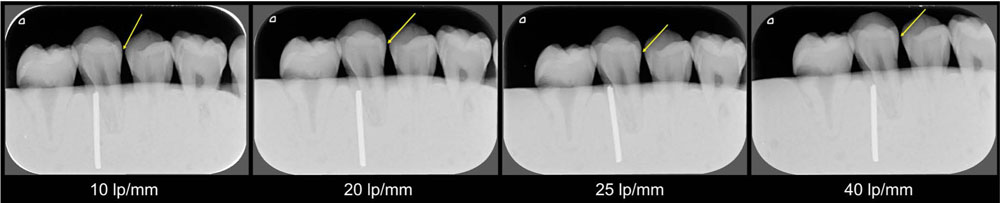
Fig. 2 Radiographic images with an enamel lesion seen in different resolutions.
Reference
-
1. Rugg-Gunn A. Dental caries: strategies to control this preventable disease. Acta Med Acad. 2013; 42:117–130.
Article2. Opal S, Garg S, Jain J, Walia I. Genetic factors affecting dental caries risk. Aust Dent J. 2015; 60:2–11.
Article3. Razmus TF. Caries, periodontal disease, and periapical changes. Dent Clin North Am. 1994; 38:13–31.4. Ferreira RI, Haiter-Neto F, Tabchoury CP, de Paiva GA, Bóscolo FN. Assessment of enamel demineralization using conventional, digital, and digitized radiography. Braz Oral Res. 2006; 20:114–119.
Article5. Wenzel A, Haiter-Neto F, Gotfredsen E. Influence of spatial resolution and bit depth on detection of small caries lesions with digital receptors. Oral Surg Oral Med Oral Pathol Oral Radiol Endod. 2007; 103:418–422.
Article6. Parks ET, Williamson GF. Digital radiography: an overview. J Contemp Dent Pract. 2002; 3:23–39.
Article7. Kalathingal SM, Shrout MK, Comer C, Brady C. Rating the extent of surface scratches on photostimulable storage phosphor plates in a dental school environment. Dentomaxillofac Radiol. 2010; 39:179–183.
Article8. Vandenberghe B, Bud M, Sutanto A, Jacobs R. The use of high-resolution digital imaging technology for small diameter K-file length determination in endodontics. Clin Oral Investig. 2010; 14:223–231.
Article9. Ludlow JB, Mol A. Digital imaging. In : White SC, Pharoah MJ, editors. Oral radiology: principles and interpretation. 7th ed. St Louis: Elsevier;2014. p. 41–62.10. Schropp L, Alyass NS, Wenzel A, Stavropoulos A. Validity of wax and acrylic as soft-tissue simulation materials used in in vitro radiographic studies. Dentomaxillofac Radiol. 2012; 41:686–690.11. Landis JR, Koch GG. The measurement of observer agreement for categorical data. Biometrics. 1977; 33:159–174.
Article12. Rocha AS, Almeida SM, Bóscolo FN, Haiter Neto F. Interexaminer agreement in caries radiographic diagnosis by conventional and digital radiographs. J Appl Oral Sci. 2005; 13:329–333.
Article13. Rockenbach MI, Veeck EB, da Costa NP. Detection of proximal caries in conventional and digital radiographs: an in vitro study. Stomatologija. 2008; 10:115–120.14. Pontual AA, de Melo DP, de Almeida SM, Bóscolo FN, Haiter Neto F. Comparison of digital systems and conventional dental film for the detection of approximal enamel caries. Dentomaxillofac Radiol. 2010; 39:431–436.
Article15. Kayipmaz S, Sezgin ÖS, Saricaoğlu ST, Çan G. An in vitro comparison of diagnostic abilities of conventional radiography, storage phosphor, and cone beam computed tomography to determine occlusal and approximal caries. Eur J Radiol. 2011; 80:478–482.
Article16. Berkhout WE, Verheij JG, Syriopoulos K, Li G, Sanderink GC, van der Stelt PF. Detection of proximal caries with high-resolution and standard resolution digital radiographic systems. Dentomaxillofac Radiol. 2007; 36:204–210.
Article17. Li G, Berkhout WE, Sanderink GC, Martins M, van der Stelt PF. Detection of in vitro proximal caries in storage phosphor plate radiographs scanned with different resolutions. Dentomaxillofac Radiol. 2008; 37:325–329.18. Nikneshan S, Abbas FM, Sabbagh S. Detection of proximal caries using digital radiographic systems with different resolutions. Indian J Dent Res. 2015; 26:5–10.
Article19. Janhom A, van Ginkel FC, van Amerongen JP, van der Stelt PF. Scanning resolution and the detection of approximal caries. Dentomaxillofac Radiol. 2001; 30:166–171.
Article20. Mistry AR, Uzbelger Feldman D, Yang J, Ryterski E. Low dose x-ray sources and high quantum efficiency sensors: the next challenge in dental digital imaging? Radiol Res Pract. 2014; 2014:543524.
Article21. Berkhout WE, Beuger DA, Sanderink GC, van der Stelt PF. The dynamic range of digital radiographic systems: dose reduction or risk of overexposure? Dentomaxillofac Radiol. 2004; 33:1–5.
Article22. Wenzel A, Møystad A. Work flow with digital intraoral radiography: a systematic review. Acta Odontol Scand. 2010; 68:106–114.
Article23. de Oliveira ML, Pinto GC, Ambrosano GM, Tosoni GM. Effect of combined digital imaging parameters on endodontic file measurements. J Endod. 2012; 38:1404–1407.
Article24. Hellén-Halme K, Lith A. Carious lesions: diagnostic accuracy using pre-calibrated monitor in various ambient light levels: an in vitro study. Dentomaxillofac Radiol. 2013; 42:20130071.
- Full Text Links
-
- Actions
-
Cited
- CITED
-
- Close
- Share
-
- Similar articles
-
- Detection method of proximal caries using line profile in digital intra-oral radiography
- Comparison of the clinical examination with the panoramic radiography in the diagnosis of dental caries
- Detection of Proximal Caries Lesions with Deep Learning Algorithm
- Proximal caries detection using digital subtraction radiography in the artificial caries activity model
- Assessment of the Object Detection Ability of Interproximal Caries on Primary Teeth in Periapical Radiographs Using Deep Learning Algorithms
