Solitary Fibrous Tumor of Breast with Anaplastic Areas (Malignant Solitary Fibrous Tumor): A Case Report with Review of Literature
- Affiliations
-
- 1Department of Pathology and Genomic Medicine, Houston Methodist Hospital, Houston, TX, USA. jaero@houstonmethodist.org
- 2Department of Pathology, Gospel Hospital, Busan, Korea.
- 3Houston Methodist Breast Surgery Partners, Baytown, TX, USA.
- 4Department of Pathology and Laboratory Medicine, Weill Medical College of Cornell University, New York, NY, USA.
- KMID: 2450125
- DOI: http://doi.org/10.4048/jbc.2019.22.e30
Abstract
- Solitary fibrous tumor (SFT) is a rare, soft tissue neoplasm that rarely presents in breast tissue, with only 27 previously reported cases. To our knowledge, only one case of malignant SFT has been reported in the English literature. A 75-year-old Caucasian woman presented to our institution with a 3-month history of a palpable left breast mass. No other symptoms, including nipple discharge or skin changes, were noted. She underwent 3 previous biopsies for right breast masses, all of which were benign, with no evidence of spindle cell neoplasm, atypical hyperplasia, or malignancy. Microscopic examination of the mass demonstrated a classic area of SFT with areas of high-grade anaplastic component. In these areas, the tumor showed atypical epithelioid cells arranged in hypercellular sheets with diminished branching vasculature, nuclear pleomorphism, and increased mitotic count (up to 9/10 high-power fields). This case represents the second case of malignant SFT in the breast.
Keyword
MeSH Terms
Figure
-
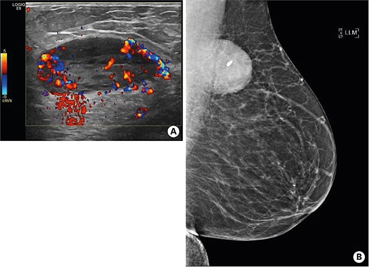
Figure 1 Radiologic findings (A) Ultrasound showing a highly vascular, oval, circumscribed, and hypoechoic mass in the left breast at posterior depth causing mass effect on the pectoralis muscle posteriorly. (B) Mammogram shows an oval, circumscribed, and obscured mass in the left breast at posterior depth abutting the pectoralis muscle. This mass contains a single benign-appearing coarse calcification.
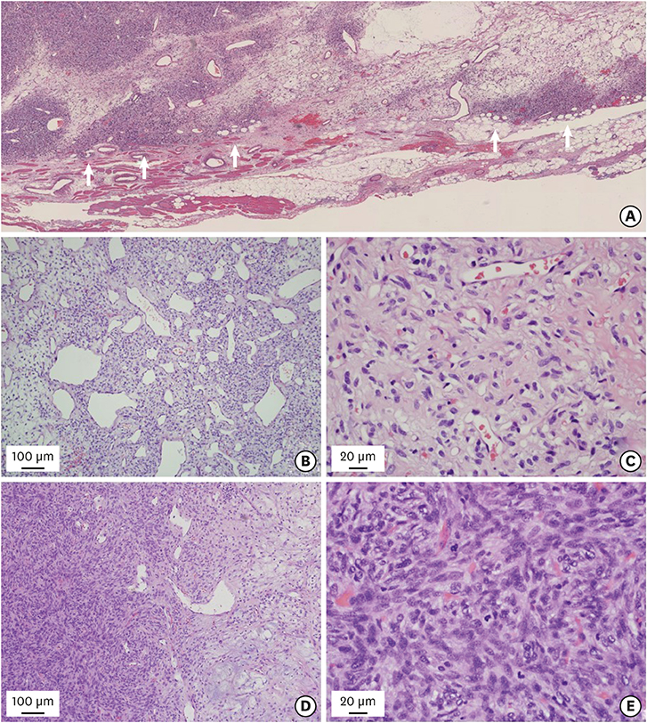
Figure 2 Microscopic findings (A) Well demarcated tumor border; however, it is focally permeative into the surrounding adipose tissue and skeletal muscles (arrows). (B) Tumor cells are arranged haphazardly with alternating cellularity (hypocellular on left and hypercellular on right) and prominent vasculature. (C) Hypocellular area is composed of bland looking, plump spindle or oval tumor cells. (D) Hypercellular area with compact sheet-like arrangement (left side) is transited from classic tumor areas (right side). (E) Hypercellular area is composed of pleomorphic tumor cells with increased mitotic figures (hematoxylin and eosin stain; (A) ×50, (B) ×100, (C) ×400, (D) ×100, and (E) ×400).
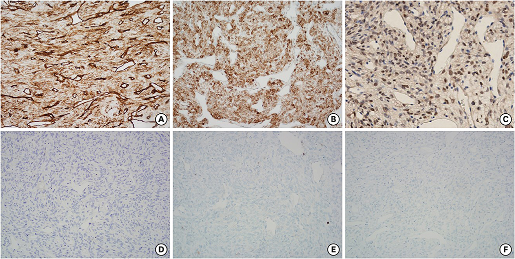
Figure 3 Tumor cells positive for cluster of differentiation 34 (A), signal transducer and activator of transcription 6 (B), B-cell lymphoma 2 (C) negative for cytokeratin (D), S100 (E), and p63 (F) (×200).

Figure 4 Immunohistochemical findings in hypercellular and pleomorphic areas cluster of differentiation 34 (A), B-cell lymphoma 2 (B), and signal transducer and activator of transcription 6 (C). Immunoreactivity shows the same staining pattern and intensity as in classic areas (×200).
Reference
-
1. Magro G, Bisceglia M, Michal M, Eusebi V. Spindle cell lipoma-like tumor, solitary fibrous tumor and myofibroblastoma of the breast: a clinico-pathological analysis of 13 cases in favor of a unifying histogenetic concept. Virchows Arch. 2002; 440:249–260.
Article2. Magro G, Sidoni A, Bisceglia M. Solitary fibrous tumour of the breast: distinction from myofibroblastoma. Histopathology. 2000; 37:189–191.
Article3. Maggiani F, Debiec-Rychter M, Verbeeck G, Sciot R. Extramammary myofibroblastoma is genetically related to spindle cell lipoma. Virchows Arch. 2006; 449:244–247.
Article4. Krismann M, Adams H, Jaworska M, Müller KM, Johnen G. Patterns of chromosomal imbalances in benign solitary fibrous tumours of the pleura. Virchows Arch. 2000; 437:248–255.
Article5. Bertucci F, Bouvier-Labit C, Finetti P, Adélaïde J, Metellus P, Mokhtari K, et al. Comprehensive genome characterization of solitary fibrous tumors using high-resolution array-based comparative genomic hybridization. Genes Chromosomes Cancer. 2013; 52:156–164.
Article6. Fritchie KJ, Carver P, Sun Y, Batiouchko G, Billings SD, Rubin BP, et al. Solitary fibrous tumor: is there a molecular relationship with cellular angiofibroma, spindle cell lipoma, and mammary-type myofibroblastoma? Am J Clin Pathol. 2012; 137:963–970.7. Magro G, Spadola S, Motta F, Palazzo J, Catalano F, Vecchio GM, et al. STAT6 expression in spindle cell lesions of the breast: an immunohistochemical study of 48 cases. Pathol Res Pract. 2018; 214:1544–1549.
Article8. Robinson DR, Wu YM, Kalyana-Sundaram S, Cao X, Lonigro RJ, Sung YS, et al. Identification of recurrent NAB2-STAT6 gene fusions in solitary fibrous tumor by integrative sequencing. Nat Genet. 2013; 45:180–185.
Article9. Yoshida A, Tsuta K, Ohno M, Yoshida M, Narita Y, Kawai A, et al. STAT6 immunohistochemistry is helpful in the diagnosis of solitary fibrous tumors. Am J Surg Pathol. 2014; 38:552–559.
Article10. Koelsche C, Schweizer L, Renner M, Warth A, Jones DT, Sahm F, et al. Nuclear relocation of STAT6 reliably predicts NAB2-STAT6 fusion for the diagnosis of solitary fibrous tumour. Histopathology. 2014; 65:613–622.
Article11. Fusco N, Guerini-Rocco E, Augello C, Terrasi A, Ercoli G, Fumagalli C, et al. Recurrent NAB2-STAT6 gene fusions and oestrogen receptor-α expression in pulmonary adenofibromas. Histopathology. 2017; 70:906–917.
Article12. Berghoff AS, Kresl P, Bienkowski M, Koelsche C, Rajky U, Hainfellner JA, et al. Validation of nuclear STAT6 immunostaining as a diagnostic marker of meningeal solitary fibrous tumor (SFT)/hemangiopericytoma. Clin Neuropathol. 2017; 36:56–59.
Article13. Yang LH, Dai SD, Li QC, Xu HT, Jiang GY, Zhang Y, et al. Malignant solitary fibrous tumor of breast: a rare case report. Int J Clin Exp Pathol. 2014; 7:4461–4466.14. Mosquera JM, Fletcher CD. Expanding the spectrum of malignant progression in solitary fibrous tumors: a study of 8 cases with a discrete anaplastic component--is this dedifferentiated SFT? Am J Surg Pathol. 2009; 33:1314–1321.
Article15. Velez-Cubian FO, Gabordi RC, Smith PV, Toloza EM. Tumor-to-tumor metastasis: an unusual case of breast cancer metastatic to a solitary fibrous tumor. J Thorac Dis. 2016; 8:E374–E378.
Article
- Full Text Links
-
- Actions
-
Cited
- CITED
-
- Close
- Share
-
- Similar articles
-
- Solitary Fibrous Tumor of the Adrenal Gland: A Case Report
- A Case of Solitary Fibrous Tumor in the Cheek
- A Case of Solitary Fibrous Tumor That Developed on the Scalp
- A Case of Solitary Fibrous Tumor of Lacrimal Gland
- Solitary fibrous tumor of the liver - An unusual entity: A case report and review of literature
