Blue Laser Imaging with a Small-Caliber Endoscope Facilitates Detection of Early Gastric Cancer
- Affiliations
-
- 1Division of Gastroenterology, Department of Medicine, Jichi Medical University, Shimotsuke, Tochigi, Japan. osawa@jichi.ac.jp
- 2Department of Surgery, Jichi Medical University, Shimotsuke, Tochigi, Japan.
- KMID: 2449769
- DOI: http://doi.org/10.5946/ce.2018.100
Abstract
- Conventional endoscopy often misses early gastric cancers with minimal red discoloration because they cannot be distinguished from inflamed mucosa. We treated a patient with a small early gastric cancer that was difficult to diagnose using conventional endoscopy. Conventional endoscopy using a small-caliber endoscope showed only subtle red discoloration of the gastric mucosa. However, blue laser imaging showed a clearly discolored area measuring 10 mm in diameter around the red lesion, which was distinct from the surrounding inflamed mucosa. Irregular vessels on the tumor surface (suspicious for early gastric cancer) were observed even with small-caliber endoscopy. Biopsy revealed a well-moderately differentiated tubular adenocarcinoma, and endoscopic submucosal dissection was performed. Histopathological examination of the specimen confirmed well-moderately differentiated adenocarcinoma localized to the mucosa with slight depression compared to the surrounding mucosa, consistent with the endoscopic findings. This small early gastric cancer became clearly visible with blue laser imaging using small-caliber endoscopy.
Keyword
MeSH Terms
Figure
-
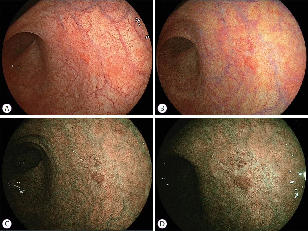
Fig. 1. (A) White light imaging with a small-caliber endoscope shows a small red area measuring 3 mm in diameter on the posterior wall near the gastric angle, which is not suspicious for gastric cancer. (B) Linked color imaging enhances the red lesion and the surrounding red portion. (C) Bright blue laser imaging reveals a discolored lesion measuring 10 mm around a central red area. (D) Blue laser imaging produces a high color contrast between the malignant lesion and the surrounding mucosa. Several irregular vessels are seen in the discolored lesion even with small-caliber endoscopy, suggesting early gastric cancer.

Fig. 2. Images obtained with a normal-caliber endoscope: (A) white light imaging and (B) linked color imaging cannot clearly reveal the site of the early gastric cancer (white arrows) because of the tangential view. (C) Blue laser imaging with middle magnification shows a brown malignant lesion surrounded by green mucosa (white arrows). (D) Blue laser imaging using high magnification shows irregular microvascular and irregular microstructural patterns on the mucosal surface.
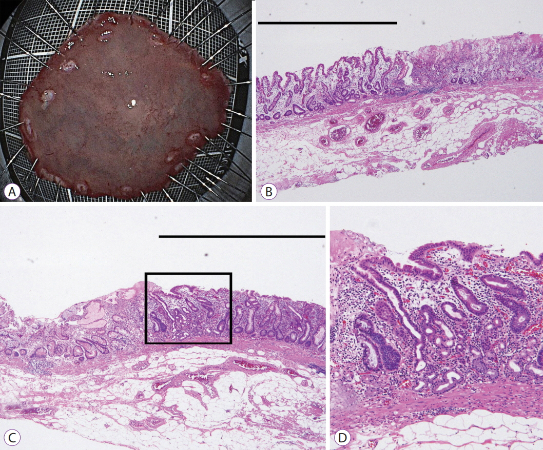
Fig. 3. (A) The specimen resected by endoscopic submucosal dissection can be observed. (B) Histopathological evaluation of the resected specimen shows well-differentiated adenocarcinoma (black line) (hematoxylin & eosin [H&E], ×40) and (C) moderately-differentiated adenocarcinoma (black line) (H&E, ×40). (D) Magnified imaging of the square in (C) shows that microvessels are present in the intervening part of the superficial layer of the mucosa (H&E, ×100).
Cited by 1 articles
-
Linked Color Imaging and Blue Laser Imaging for Upper Gastrointestinal Screening
Hiroyuki Osawa, Yoshimasa Miura, Takahito Takezawa, Yuji Ino, Tsevelnorov Khurelbaatar, Yuichi Sagara, Alan Kawarai Lefor, Hironori Yamamoto
Clin Endosc. 2018;51(6):513-526. doi: 10.5946/ce.2018.132.
Reference
-
1. Raftopoulos SC, Segarajasingam DS, Burke V, Ee HC, Yusoff IF. A cohort study of missed and new cancers after esophagogastroduodenoscopy. Am J Gastroenterol. 2010; 105:1292–1297.
Article2. Fukuda H, Miura Y, Hayashi Y, et al. Linked color imaging technology facilitates early detection of flat gastric cancers. Clin J Gastroenterol. 2015; 8:385–389.
Article3. Yoshida N, Yagi N, Inada Y, et al. Ability of a novel blue laser imaging system for the diagnosis of colorectal polyps. Dig Endosc. 2014; 26:250–258.
Article4. Okada M, Sakamoto H, Takezawa T, et al. Laterally spreading tumor of the rectum delineated with linked color imaging technology. Clin Endosc. 2016; 49:207–208.
Article5. Dohi O, Yagi N, Onozawa Y, et al. Linked color imaging improves endoscopic diagnosis of active Helicobacter pylori infection. Endosc Int Open. 2016; 4:E800–E805.
Article6. Osawa H, Yamamoto H, Miura Y, et al. Blue laser imaging provides excellent endoscopic images of upper gastrointestinal lesions. Video Journal and Encyclopedia of GI Endoscopy. 2014; 1:607–610.
Article7. Osawa H, Yamamoto H. Present and future status of flexible spectral imaging color enhancement and blue laser imaging technology. Dig Endosc. 2014; 26 Suppl 1:105–115.
Article8. Toma S, Osawa H, Yoshizawa M, et al. Diagnosis of small flat early gastric cancer by flexible spectral imaging color enhancement. Clin J Gastroenterol. 2010; 3:88–91.
Article9. Zaman A, Hahn M, Hapke R, Knigge K, Fennerty MB, Katon RM. A randomized trial of peroral versus transnasal unsedated endoscopy using an ultrathin videoendoscope. Gastrointest Endosc. 1999; 49(3 Pt 1):279–284.
Article10. Birkner B, Fritz N, Schatke W, Hasford J. A prospective randomized comparison of unsedated ultrathin versus standard esophagogastroduodenoscopy in routine outpatient gastroenterology practice: does it work better through the nose? Endoscopy. 2003; 35:647–651.
Article11. Faulx AL, Catanzaro A, Zyzanski S, et al. Patient tolerance and acceptance of unsedated ultrathin esophagoscopy. Gastrointest Endosc. 2002; 55:620–623.
Article12. Mori M, Adachi Y, Kakeji Y, et al. Superficial flat-type early carcinoma of the stomach. Cancer. 1992; 69:306–313.
Article13. Nakamura K, Sugano H, Takagi K. Carcinoma of the stomach in incipient phase: its histogenesis and histological appearances. Gan. 1968; 59:251–258.14. Matsukura N, Suzuki K, Kawachi T, et al. Distribution of marker enzymes and mucin in intestinal metaplasia in human stomach and relation to complete and incomplete types of intestinal metaplasia to minute gastric carcinomas. J Natl Cancer Inst. 1980; 65:231–240.15. Kohli Y, Pfeiffer CJ, Kutty KP, Barrowman JA, Heughan C, Kepkay DL. Endoscopic diagnosis of intestinal metaplasia in Canada and Japan. J Clin Gastroenterol. 1981; 3 Suppl 1:29–33.
Article16. Yoshida N, Hisabe T, Inada Y, et al. The ability of a novel blue laser imaging system for the diagnosis of invasion depth of colorectal neoplasms. J Gastroenterol. 2014; 49:73–80.
Article17. Dohi O, Yagi N, Majima A, et al. Diagnostic ability of magnifying endoscopy with blue laser imaging for early gastric cancer: a prospective study. Gastric Cancer. 2017; 20:297–303.
Article18. Ikematsu H, Sakamoto T, Togashi K, et al. Detectability of colorectal neoplastic lesions using a novel endoscopic system with blue laser imaging: a multicenter randomized controlled trial. Gastrointest Endosc. 2017; 86:386–394.
Article19. Tomie A, Dohi O, Yagi N, et al. Blue laser imaging-bright improves endoscopic recognition of superficial esophageal squamous cell carcinoma. Gastroenterol Res Pract. 2016; 2016:6140854.
Article20. Iwashita C, Miura Y, Osawa H, et al. Laser imaging facilitates early detection of synchronous adenocarcinomas in patients with Barrett’s esophagus. Clin Endosc. 2017; 50:81–86.
Article
- Full Text Links
-
- Actions
-
Cited
- CITED
-
- Close
- Share
-
- Similar articles
-
- Linked Color Imaging and Blue Laser Imaging for Upper Gastrointestinal Screening
- Laser Imaging Facilitates Early Detection of Synchronous Adenocarcinomas in Patients with Barrett's Esophagus
- Appropriate Color Enhancement Settings for Blue Laser Imaging Facilitates the Diagnosis of Early Gastric Cancer with High Color Contrast
- Detection of Gastrointestinal Cancer using Linked Color Imaging and Blue Light Imaging
- Clinical Applications of Linked Color Imaging and Blue Laser/Light Imaging in the Screening, Diagnosis, and Treatment of Superficial Colorectal Tumors
