Bimodal Chromoendoscopy with Confocal Laser Endomicroscopy for the Detection of Early Esophageal Squamous Cell Neoplasms
- Affiliations
-
- 1Division of Gastroenterology, Department of Medicine, Faculty of Medicine, Chulalongkorn University and King Chulalongkorn Memorial Hospital, Bangkok, Thailand. ercp@live.com
- 2Division of General Internal Medicine, Department of Medicine, Faculty of Medicine, Chulalongkorn University and King Chulalongkorn Memorial Hospital, Bangkok, Thailand.
- 3Department of Pathology, Faculty of Medicine, Chulalongkorn University and King Chulalongkorn Memorial Hospital, Bangkok, Thailand.
- KMID: 2447671
- DOI: http://doi.org/10.5946/ce.2018.091
Abstract
- BACKGROUND/AIMS
This study aimed to evaluate the diagnostic accuracy of dual-focus narrow-band imaging (dNBI) and Lugol'schromoendoscopy (LCE) combined with probe-based confocal laser endomicroscopy (pCLE) to screen for esophageal squamous cell neoplasms (ESCNs) in patients with a history of head and neck cancer.
METHODS
From March to August 2016, dNBI was performed. Next, LCE was performed, followed by pCLE and biopsy. Histology has historically been the gold standard to diagnose ESCN. The sensitivity, specificity, positive predictive value (PPV), negative predictive value (NPV), and accuracy of dNBI and LCE adjunct with pCLE were determined.
RESULTS
Twenty-four patients were included. Ten ESCNs were found in 8 patients (33%). Forty percent of high-graded intraepithelial neoplasias and all low-grade intraepithelial neoplasias were overlooked by dNBI. The sensitivity, specificity, PPV, NPV, and accuracy of dNBI vs. LCE combined with pCLE were 50% vs. 80%, 62% vs. 67%, 36% vs. 44%, 75% vs. 91%, and 83% vs. 70%, respectively.
CONCLUSIONS
The use of dNBI to detect ESCN was suboptimal. LCE with pCLE following dNBI had additional value for detecting esophageal dysplasia not detected by dNBI. The use of pCLE to detect dNBI-missed lesions yielded a high NPV, while pCLE-guided biopsy could reduce the number of unnecessary biopsies.
Keyword
MeSH Terms
Figure
-
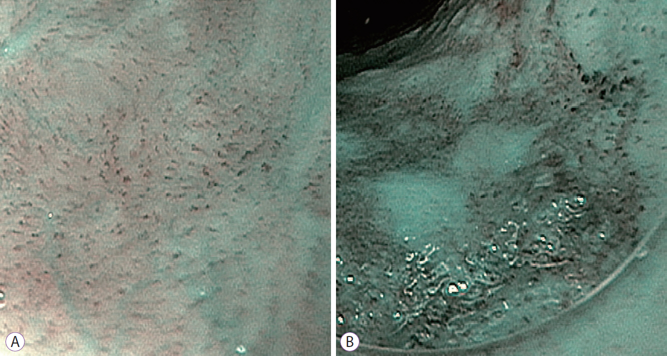
Fig. 1. Near focus view of dual-focus narrow-band imaging (dNBI) of normal esophageal mucosa showing a normal intrapapillary capillary loop (IPCL) pattern (A). The near focus view of dNBI of high-grade intraepithelial neoplasia shows a well-demarcated brownish area with dilated, tortuous, and various shapes of IPCL (B).
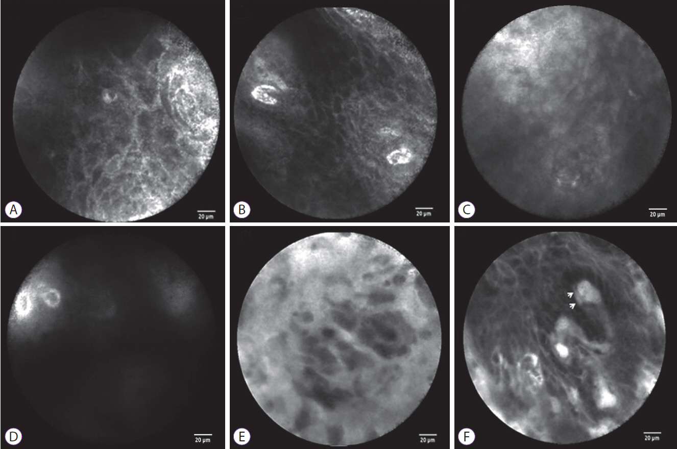
Fig. 2. The probe-based confocal laser endomicroscopy (pCLE) findings of normal esophageal mucosa and esophageal squamous cell neoplasms. For normal esophageal mucosa, pCLE shows a homogenous, regular architecture, clearly visible cell borders (A), and regular capillaries without fluorescein leakage (B). For low-grade intraepithelial neoplasia, pCLE shows a slightly inhomogeneous, irregular architecture, clearly visible cell borders (C), and regular capillaries without fluorescein leakage (D). For high-grade intraepithelial neoplasia, pCLE shows an inhomogeneous and irregular cellular architecture without clearly visible cell borders (E) and irregular, twisted, dilated, elongated capillaries (F) with fluorescein leakage (F, arrows).
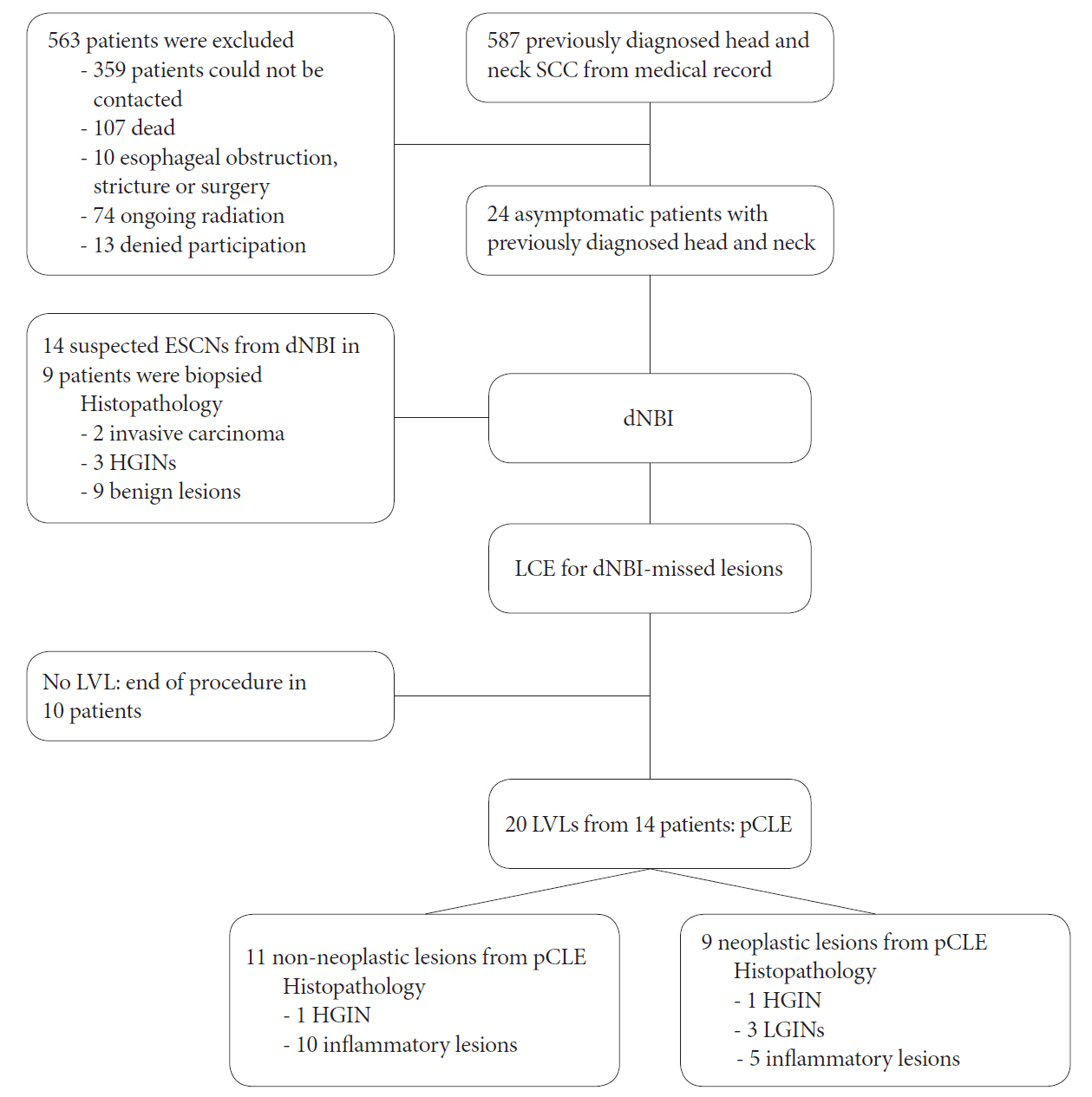
Fig. 3. Flow chart of the studied patients. dNBI, dual-focus narrow-band imaging; ESCN, esophageal squamous cell neoplasm; HGIN, high-grade intraepithelial neoplasia; LCE, Lugol’s chromoendoscopy; LGIN, low-grade intraepithelial neoplasia; LVL, Lugol’s voiding lesion; pCLE, probe-based confocal laser endomicroscopy; SCC, squamous cell carcinoma.
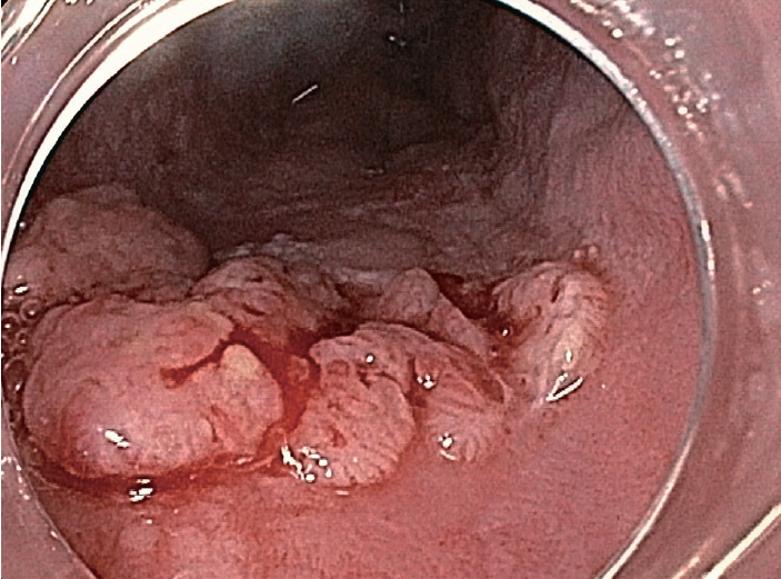
Fig. 4. Squamous cell cancer detected by white-light endoscopy showing a nodular surface with dilated neoplastic vessels.
Cited by 1 articles
-
Usefulness of Probe-Based Confocal Laser Endomicroscopy for Esophageal Squamous Cell Neoplasm
Sang Kil Lee
Clin Endosc. 2019;52(2):91-92. doi: 10.5946/ce.2019.054.
Reference
-
1. Jemal A, Bray F, Center MM, Ferlay J, Ward E, Forman D. Global cancer statistics. CA Cancer J Clin. 2011; 61:69–90.
Article2. Wang GQ, Jiao GG, Chang FB, et al. Long-term results of operation for 420 patients with early squamous cell esophageal carcinoma discovered by screening. Ann Thorac Surg. 2004; 77:1740–1744.
Article3. Rustgi AK, El-Serag HB. Esophageal carcinoma. N Engl J Med. 2014; 371:2499–2509.
Article4. Katada C, Muto M, Momma K, et al. Clinical outcome after endoscopic mucosal resection for esophageal squamous cell carcinoma invading the muscularis mucosae--a multicenter retrospective cohort study. Endoscopy. 2007; 39:779–783.5. Hirota WK, Zuckerman MJ, Adler DG, et al. ASGE guideline: the role of endoscopy in the surveillance of premalignant conditions of the upper GI tract. Gastrointest Endosc. 2006; 63:570–580.
Article6. Freitag CP, Barros SG, Kruel CD, et al. Esophageal dysplasias are detected by endoscopy with Lugol in patients at risk for squamous cell carcinoma in southern Brazil. Dis Esophagus. 1999; 12:191–195.
Article7. Prueksapanich P, Pittayanon R, Rerknimitr R, Wisedopas N, Kullavanijaya P. Value of probe-based confocal laser endomicroscopy (pCLE) and dual focus narrow-band imaging (dNBI) in diagnosing early squamous cell neoplasms in esophageal Lugol’s voiding lesions. Endosc Int Open. 2015; 3:E281–E288.
Article8. Lee CT, Chang CY, Lee YC, et al. Narrow-band imaging with magnifying endoscopy for the screening of esophageal cancer in patients with primary head and neck cancers. Endoscopy. 2010; 42:613–619.
Article9. Takenaka R, Kawahara Y, Okada H, et al. Narrow-band imaging provides reliable screening for esophageal malignancy in patients with head and neck cancers. Am J Gastroenterol. 2009; 104:2942–2948.
Article10. Nagami Y, Tominaga K, Machida H, et al. Usefulness of non-magnifying narrow-band imaging in screening of early esophageal squamous cell carcinoma: a prospective comparative study using propensity score matching. Am J Gastroenterol. 2014; 109:845–854.
Article11. Chen MC, Feng IJ, Lu CH, et al. The incidence and risk of second primary cancers in patients with nasopharyngeal carcinoma: a population-based study in Taiwan over a 25-year period (1979-2003). Ann Oncol. 2008; 19:1180–1186.
Article12. Inoue H, Kaga M, Ikeda H, et al. Magnification endoscopy in esophageal squamous cell carcinoma: a review of the intrapapillary capillary loop classification. Ann Gastroenterol. 2015; 28:41–48.13. Hashimoto CL, Iriya K, Baba ER, et al. Lugol’s dye spray chromoendoscopy establishes early diagnosis of esophageal cancer in patients with primary head and neck cancer. Am J Gastroenterol. 2005; 100:275–282.
Article14. Liu H, Li YQ, Yu T, et al. Confocal laser endomicroscopy for superficial esophageal squamous cell carcinoma. Endoscopy. 2009; 41:99–106.
Article15. Lee YC, Wang CP, Chen CC, et al. Transnasal endoscopy with narrow-band imaging and Lugol staining to screen patients with head and neck cancer whose condition limits oral intubation with standard endoscope (with video). Gastrointest Endosc. 2009; 69:408–417.
Article16. Day GL, Blot WJ. Second primary tumors in patients with oral cancer. Cancer. 1992; 70:14–19.
Article17. Priante AV, Castilho EC, Kowalski LP. Second primary tumors in patients with head and neck cancer. Curr Oncol Rep. 2011; 13:132–137.
Article18. Schwartz LH, Ozsahin M, Zhang GN, et al. Synchronous and metachronous head and neck carcinomas. Cancer. 1994; 74:1933–1938.
Article19. Goda K, Dobashi A, Yoshimura N, et al. Dual-focus versus conventional magnification endoscopy for the diagnosis of superficial squamous neoplasms in the pharynx and esophagus: a randomized trial. Endoscopy. 2016; 48:321–329.
Article20. Dawsey SM, Lewin KJ, Wang GQ, et al. Squamous esophageal histology and subsequent risk of squamous cell carcinoma of the esophagus. A prospective follow-up study from Linxian, China. Cancer. 1994; 74:1686–1692.21. Su YY, Chen WC, Chuang HC, et al. Effect of routine esophageal screening in patients with head and neck cancer. JAMA Otolaryngol Head Neck Surg. 2013; 139:350–354.
Article22. Li J, Xu R, Liu M, et al. Lugol chromoendoscopy detects esophageal dysplasia with low levels of sensitivity in a high-risk region of China. Clin Gastroenterol Hepatol. 2018; 16:1585–1592.
Article
- Full Text Links
-
- Actions
-
Cited
- CITED
-
- Close
- Share
-
- Similar articles
-
- Usefulness of Probe-Based Confocal Laser Endomicroscopy for Esophageal Squamous Cell Neoplasm
- Confocal Laser Endomicroscopy and Molecular Imaging in Barrett Esophagus and Stomach
- Role of Advanced Endoscopic Imaging Techniques in the Management of Inflammatory Bowel Disease
- Diagnosis and Clinical Management of Esophageal Squamous Dysplasia
- Usefulness and Future Prospects of Confocal Laser Endomicroscopy for Gastric Premalignant and Malignant Lesions
