Neurointervention.
2019 Mar;14(1):53-60. 10.5469/neuroint.2019.00024.
Efficacy of Percutaneous Sclerotherapy in Low Flow Venous Malformations - A Single Center Series
- Affiliations
-
- 1Department of Neuroradiology, Lahore General Hospital, Lahore, Pakistan. masterinfluencer@gmail.com
- KMID: 2446782
- DOI: http://doi.org/10.5469/neuroint.2019.00024
Abstract
- PURPOSE
We analyzed results of percutaneous sclerotherapy for venous malformations (VMs) in head, neck and extremities.
MATERIALS AND METHODS
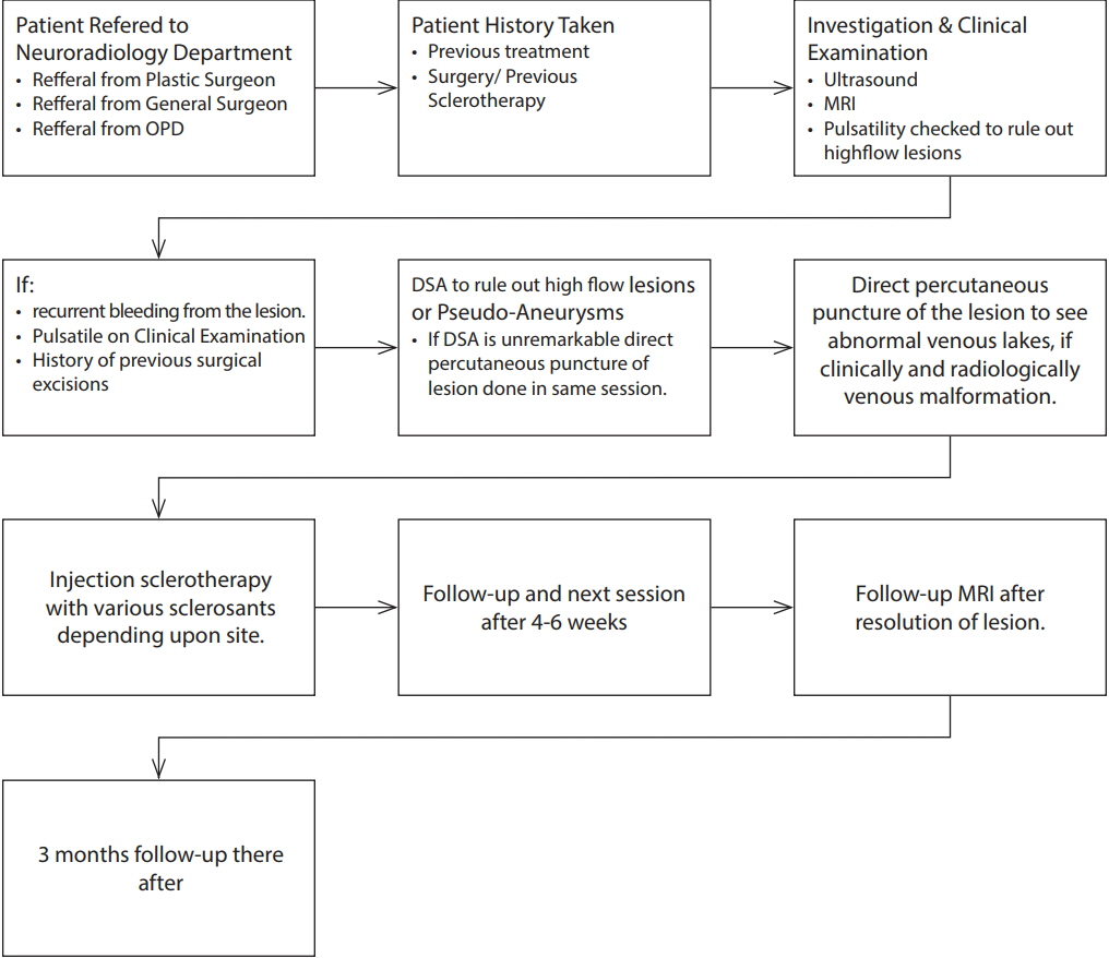
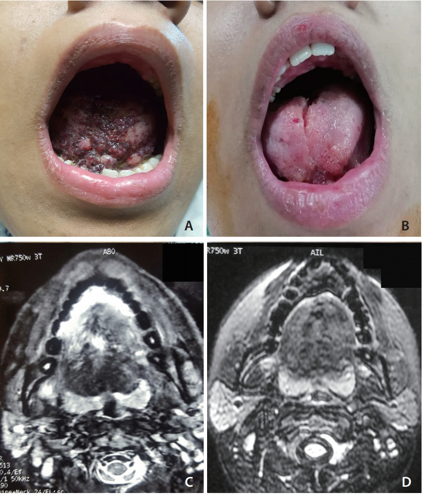
Thirty-five patients with head and neck and extremities VM treated by sclerotherapy with bleomycin and sodium tetradecyl sulphate (STS) were retrospectively reviewed. A pre-treatment magnetic resonance imaging was done for all patients to diagnose the lesion. Each lesion received 1 to 11 sessions (average, 2.7; standard deviation [SD], 2.03). We evaluated percentage reduction in swelling size and a Likert scale review of subjective feelings of the patients.
RESULTS
Sixteen had a complete obliteration; by sclerotherapy alone (n=13) and surgery after a 75% reduction (n=3). Ten patients had a significant reduction up to 75% and three patients by 50%. Four had a minimal decrease with reduction of 25% or less. Follow-up duration of the patients varied from a minimum of 6 months up to 3 years (average, 15.7 months; SD, 7.8 months). Of all patients, three refused further treatment and were lost to follow-up, while another two were referred to a dermatologist. Thirteen patients reported feeling excellent after the sessions. Eight patients claimed to feel slightly better compared to before the sessions started. Only three patients complained of feeling the same before and after the sessions. None of the patients still in follow-up have reported a recurrence of a lesion thus far.
CONCLUSION
Sclerotherapy using bleomycin and STS as sclerosants is a safe and effective primary treatment for VMs in the head and neck as well as in extremities.
MeSH Terms
Figure
Cited by 1 articles
-
Commentary to: Efficacy of Percutaneous Sclerotherapy in Low Flow Venous Malformations - A Single Center Series
Jongmin Lee
Neurointervention. 2019;14(1):61-62. doi: 10.5469/neuroint.2019.00059.
Reference
-
1. Behravesh S, Yakes W, Gupta N, Naidu S, Chong BW, Khademhosseini A, et al. Venous malformations: clinical diagnosis and treatment. Cardiovasc Diagn Ther. 2016; 6:557–569.
Article2. Spence J, Krings T, terBrugge KG, da Costa LB, Agid R. Percutaneous sclerotherapy for facial venous malformations: subjective clinical and objective MR imaging follow-up results. AJNR Am J Neuroradiol. 2010; 31:955–960.
Article3. Dubois J, Soulez G, Oliva VL, Berthiaume MJ, Lapierre C, Therasse E. Soft-tissue venous malformations in adult patients: imaging and therapeutic issues. Radiographics. 2001; 21:1519–1531.
Article4. Rautio R, Laranne J, Kähärä V, Saarinen J, Keski-Nisula L. Long-term results and quality of life after endovascular treatment of venous malformations in the face and neck. Acta Radiol. 2004; 45:738–745.
Article5. Mathur NN, Rana I, Bothra R, Dhawan R, Kathuria G, Pradhan T. Bleomycin sclerotherapy in congenital lymphatic and vascular malformations of head and neck. Int J Pediatr Otorhinolaryngol. 2005; 69:75–80.
Article6. Bajpai H, Bajpai S. Comparative analysis of intralesional sclerotherapy with sodium tetradecyl sulfate versus bleomycin in the management of low flow craniofacial soft tissue vascular lesions. J Maxillofac Oral Surg. 2012; 11:13–20.
Article7. Horbach SE, Lokhorst MM, Saeed P, de Goüyon Matignon de Pontouraude CM, Rothová A, van der Horst CM. Sclerotherapy for low-flow vascular malformations of the head and neck: a systematic review of sclerosing agents. J Plast Reconstr Aesthet Surg. 2016; 69:295–304.8. Bashir U, Shah S, Jeph S, O’Keeffe M, Khosa F. Magnetic resonance (MR) imaging of vascular malformations. Pol J Radiol. 2017; 82:731–741.
Article9. Balasundaram I, Al-Hadad I, Rehman K, McCafferty I, Monaghan A. The use of foam sclerotherapy to treat low-flow vascular malformations of the head and neck. J Surg Case Rep. 2014; 2014:rju095.
Article10. Alakailly X, Kummoona R, Quereshy FA, Baur DA, González AE. The use of sodium tetradecyl sulphate for the treatment of venous malformations of the head and neck. J Maxillofac Oral Surg. 2015; 14:332–328.
Article11. Konez O, Burrows PE. Magnetic resonance of vascular anomalies. Magn Reson Imaging Clin N Am. 2002; 10:363–388. vii.
Article12. El-Merhi F, Garg D, Cura M, Ghaith O. Peripheral vascular tumors and vascular malformations: imaging (magnetic resonance imaging and conventional angiography), pathologic correlation and treatment options. Int J Cardiovasc Imaging. 2013; 29:379–393.
Article13. Griffiths TL, Burr ML, Campbell IA, Lewis-Jenkins V, Mullins J, Shiels K, et al. Results at 1 year of outpatient multidisciplinary pulmonary rehabilitation: a randomised controlled trial. Lancet. 2000; 355:362–368.
Article14. Muir T, Kirsten M, Fourie P, Dippenaar N, Ionescu GO. Intralesional bleomycin injection (IBI) treatment for haemangiomas and congenital vascular malformations. Pediatr Surg Int. 2004; 19:766–773.
Article
- Full Text Links
- Actions
-
Cited
- CITED
-
- Close
- Share
- Similar articles
-
- Commentary to: Efficacy of Percutaneous Sclerotherapy in Low Flow Venous Malformations - A Single Center Series
- Therapeutic Effect of Sclerotherapy on Venous Malformations
- Hemangioma and Arteriovenous Malformation: Percutaneous Sclerotherapy and Endovascular Treatment
- Sclerotherapy for Venous Malformations of Head and Neck: Systematic Review and Meta-Analysis
- A Case of Superior Mesenteric Venous Thrombosis after Endoscopic Sclerotherapy


