Ann Dermatol.
2019 Jun;31(3):343-345. 10.5021/ad.2019.31.3.343.
Isolated Phthiriasis Palpebrarum in an Elderly Woman: Diagnosis and Treatment Using by Dermoscopy
- Affiliations
-
- 1Department of Dermatology, Chonbuk National University Medical School, Jeonju, Korea. airmd@jbnu.ac.kr
- 2Department of Internal Medicine, Chonbuk National University Medical School, Jeonju, Korea.
- 3Research Institute of Clinical Medicine of Chonbuk National University-Biomedical Research Institute of Chonbuk National University Hospital, Jeonju, Korea.
- KMID: 2444871
- DOI: http://doi.org/10.5021/ad.2019.31.3.343
Abstract
- No abstract available.
MeSH Terms
Figure
-
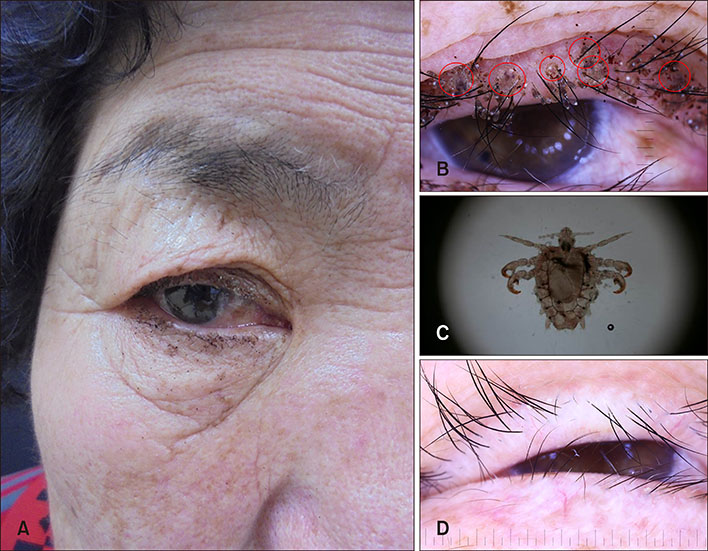
Fig. 1 (A) Naked eye examination showed erythema and multiple black granules around the right eyelid. (B) Dermoscopy revealed several crab lice (circles), ovoid nits, and red-brown feces on the right eyelash and eyelid (×30). (C) Pthirus pubis was removed from the eyelash (×100). (D) Dermoscopy of the eyelid revealed complete clearance without lice or nits after 4 weeks of follow-up (×30).
Reference
-
1. Ko CJ, Elston DM. Pediculosis. J Am Acad Dermatol. 2004; 50:1–12.
Article2. Padhi TR, Das S, Sharma S, Rath S, Rath S, Tripathy D, et al. Ocular parasitoses: a comprehensive review. Surv Ophthalmol. 2017; 62:161–189.
Article3. Turgut B, Kurt J, Catak O, Demir T. Phthriasis palpebrarum mimicking lid eczema and blepharitis. J Ophthalmol. 2009; 2009:803951.
Article4. Micali G, Lacarrubba F, Massimino D, Schwartz RA. Dermatoscopy: alternative uses in daily clinical practice. J Am Acad Dermatol. 2011; 64:1135–1146.
Article5. DeFazio JL, Spencer P. Images in clinical medicine. Dermoscopy of phthiriasis. N Engl J Med. 2010; 362:e33.
