Comparison of the accuracy of intraoral scanner by three-dimensional analysis in single and 3-unit bridge abutment model: In vitro study
- Affiliations
-
- 1Department of Prosthodontics, School of Dentistry, Kyungpook National University, Daegu, Republic of Korea. kblee@knu.ac.kr
- 2Department of Dental Science, Graduate School, Kyungpook National University, Daegu, Republic of Korea.
- 3Advanced Dental Device Development Institute (A3DI), Kyungpook National University, Daegu, Republic of Korea.
- KMID: 2444178
- DOI: http://doi.org/10.4047/jkap.2019.57.2.102
Abstract
- PURPOSE
The purpose of this study was to evaluate the accuracy of three types of intraoral scanners and the accuracy of the single abutment and bridge abutment model.
MATERIALS AND METHODS
In this study, a single abutment, and a bridge abutment with missing first molar was fabricated and set as the reference model. The reference model was scanned with an industrial three-dimensional scanner and set as reference scan data. The reference model was scanned five times using the three intraoral scanners (CS3600, CS3500, and EZIS PO). This was set as the evaluation scan data. In the three-dimensional analysis (Geomagic control X), the divided abutment region was selected and analyzed to verify the scan accuracy of the abutment. Statistical analysis was performed using SPSS software (α = .05). The accuracy of intraoral scanners was compared using the Kruskal-Wallis test and post-test was performed using the Pairwise test. The accuracy difference between the single abutment model and the bridge abutment model was analyzed by the Mann-Whitney U test.
RESULTS
The accuracy according to the intraoral scanner was significantly different (P < .05). The trueness of the single abutment model and the bridge abutment model showed a statistically significant difference and showed better trueness in the single abutment (P < .05). There was no significant difference in the precision (P = .616).
CONCLUSION
As a result of comparing the accuracy of single and bridge abutments, the error of abutment scan increased with increasing scan area, and the accuracy of bridge abutment model was clinically acceptable in three types of intraoral scanners.
MeSH Terms
Figure
-
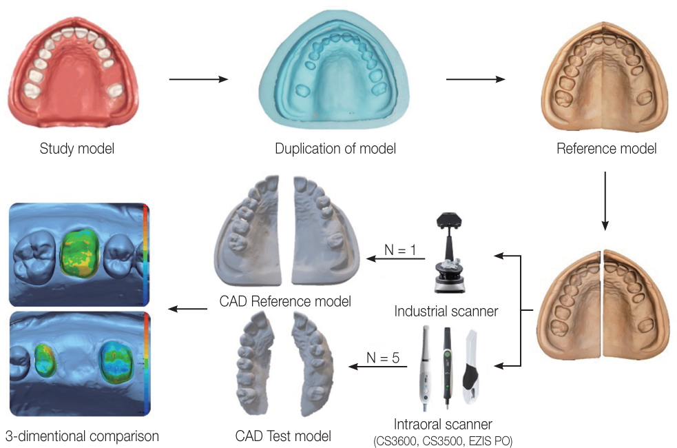
Fig. 1 Experimental design.
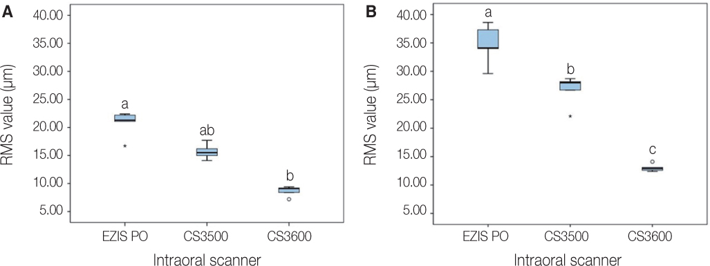
Fig. 2 Comparison of trueness according to intraoral scanner. (A) Single abutment, (B) Bridge abutment. Different letters indicate significant differences (P < .05). Circle points indicate outliers.
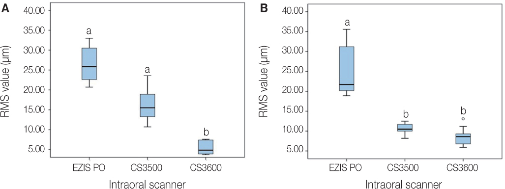
Fig. 3 Comparison of precision according to intraoral scanner. (A) Single abutment, (B) Bridge abutment. Different letters indicate significant differences (P < .05). Circle points indicate outliers.
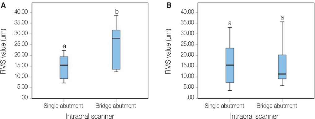
Fig. 4 Comparison of accuracy according to abutment type. (A) Trueness, (B) Precision. Different letters indicate significant differences (P < .05).
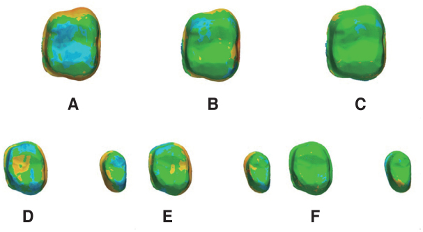
Fig. 5 Comparison of color difference map according to intraoral scanner. (A) EZIS PO (single), (B) CS3500 (single), (C) CS3600 (single), (D) EZIS PO (bridge), (E) CS3500 (bridge), (F) CS3600 (bridge).
Reference
-
1. Ng J, Ruse D, Wyatt C. A comparison of the marginal fit of crowns fabricated with digital and conventional methods. J Prosthet Dent. 2014; 112:555–560.
Article2. Wesemann C, Muallah J, Mah J, Bumann A. Accuracy and efficiency of full-arch digitalization and 3D printing: A comparison between desktop model scanners, an intraoral scanner, a CBCT model scan, and stereolithographic 3D printing. Quintessence Int. 2017; 48:41–50.3. Kang BG, Kim HJ, Chung CH. Accuracy of the CT guided implant template by using an intraoral scanner according to the edentulous distance. J Korean Acad Prosthodont. 2017; 55:1–8.
Article4. Sedda M, Casarotto A, Raustia A, Borracchini A. Effect of storage time on the accuracy of casts made from different irreversible hydrocolloids. J Contemp Dent Pract. 2008; 9:59–66.
Article5. Chandran DT, Jagger DC, Jagger RG, Barbour ME. Two- and three-dimensional accuracy of dental impression materials: effects of storage time and moisture contamination. Biomed Mater Eng. 2010; 20:243–249.
Article6. Mah J, Hatcher D. Current status and future needs in craniofacial imaging. Orthod Craniofac Res. 2003; 6:10–16.
Article7. White AJ, Fallis DW, Vandewalle KS. Analysis of intraarch and interarch measurements from digital models with 2 impression materials and a modeling process based on cone-beam computed tomography. Am J Orthod Dentofacial Orthop. 2010; 137:456.e1–456.e9.
Article8. Kwon HJ, Kim KK, Yi WJ. Comparison of digital models generated from three-dimensional optical scanner and cone beam computed tomography. J Dent Rehabil Appl Sci. 2016; 32:60–69.
Article9. Persson A, Andersson M, Oden A, Sandborgh-Englund G. A three-dimensional evaluation of a laser scanner and a touchprobe scanner. J Prosthet Dent. 2006; 95:194–200.
Article10. van Noort R. The future of dental devices is digital. Dent Mater. 2012; 28:3–12.
Article11. Park JH, Seol JH, Lee JJ, Lee SP, Lim YJ. Comparative study on quality of scanned images from varying materials and surface conditions of standardized model for dental scanner evaluation. J Dent Rehabil Appl Sci. 2018; 34:104–115.
Article12. Felton DA, Kanoy BE, Bayne SC, Wirthman GP. Effect of in vivo crown margin discrepancies on periodontal health. J Prosthet Dent. 1991; 65:357–364.
Article13. ISO 5725-1. Accuracy (trueness and precision) of measurement methods and results - Part 1: General principles and definitions. Geneva; Switzerland: International Standards Organization (ISO);1994. Available at: https://www.iso.org/obp/ui/#iso:std:iso:5725:-1:ed-1:v1:en.14. Lee JJ, Jeong ID, Park JY, Jeon JH, Kim JH, Kim WC. Accuracy of single-abutment digital cast obtained using intraoral and cast scanners. J Prosthet Dent. 2017; 117:253–259.
Article15. Bohner LOL, De Luca Canto G, Marció BS, Laganá DC, Sesma N, Tortamano Neto P. Computer-aided analysis of digital dental impressions obtained from intraoral and extraoral scanners. J Prosthet Dent. 2017; 118:617–623.
Article16. Vecsei B, Joós-Kovács G, Borbély J, Hermann P. Comparison of the accuracy of direct and indirect three-dimensional digitizing processes for CAD/CAM systems - An in vitro study. J Prosthodont Res. 2017; 61:177–184.
Article17. Patzelt SB, Emmanouilidi A, Stampf S, Strub JR, Att W. Accuracy of full-arch scans using intraoral scanners. Clin Oral Investig. 2014; 18:1687–1694.
Article18. Ender A, Mehl A. In-vitro evaluation of the accuracy of conventional and digital methods of obtaining full-arch dental impressions. Quintessence Int. 2015; 46:9–17.19. Ender A, Mehl A. Full arch scans: conventional versus digital impressions-an in-vitro study. Int J Comput Dent. 2011; 14:11–21.20. Ender A, Mehl A. Accuracy of complete-arch dental impressions: a new method of measuring trueness and precision. J Prosthet Dent. 2013; 109:121–128.
Article21. Jeong ID, Lee JJ, Jeon JH, Kim JH, Kim HY, Kim WC. Accuracy of complete-arch model using an intraoral video scanner: An in vitro study. J Prosthet Dent. 2016; 115:755–759.
Article22. Flügge TV, Schlager S, Nelson K, Nahles S, Metzger MC. Precision of intraoral digital dental impressions with iTero and extraoral digitization with the iTero and a model scanner. Am J Orthod Dentofacial Orthop. 2013; 144:471–478.
Article23. Kim KR, Seo K, Kim S. Comparison of the accuracy of digital impressions and traditional impressions: Systematic review. J Korean Acad Prosthodont. 2018; 56:258–268.
Article24. Ender A, Attin T, Mehl A. In vivo precision of conventional and digital methods of obtaining complete-arch dental impressions. J Prosthet Dent. 2016; 115:313–320.
Article25. Grünheid T, McCarthy SD, Larson BE. Clinical use of a direct chairside oral scanner: an assessment of accuracy, time, and patient acceptance. Am J Orthod Dentofacial Orthop. 2014; 146:673–682.
Article26. Aragón ML, Pontes LF, Bichara LM, Flores-Mir C, Normando D. Validity and reliability of intraoral scanners compared to conventional gypsum models measurements: a systematic review. Eur J Orthod. 2016; 38:429–434.
Article27. Choi JH, Lim YJ, Lee WJ, Han JS, Lee SP. Review of recent developments for intra-oral scanners. J Dent Rehabil Appl Sci. 2015; 31:112–125.
Article28. Kim J, Park JM, Kim M, Heo SJ, Shin IH, Kim M. Comparison of experience curves between two 3-dimensional intraoral scanners. J Prosthet Dent. 2016; 116:221–230.
Article29. Son K, Lee WS, Lee KB. Prediction of the learning curves of 2 dental CAD software programs. J Prosthet Dent. 2019; 121:95–100.
Article30. Kim RJ, Park JM, Shim JS. Accuracy of 9 intraoral scanners for complete-arch image acquisition: A qualitative and quantitative evaluation. J Prosthet Dent. 2018; 120:895–903.
Article31. Imburgia M, Logozzo S, Hauschild U, Veronesi G, Mangano C, Mangano FG. Accuracy of four intraoral scanners in oral implantology: a comparative in vitro study. BMC Oral Health. 2017; 17:92.
Article32. Fukazawa S, Odaira C, Kondo H. Investigation of accuracy and reproducibility of abutment position by intraoral scanners. J Prosthodont Res. 2017; 61:450–459.
Article33. Nedelcu RG, Persson AS. Scanning accuracy and precision in 4 intraoral scanners: an in vitro comparison based on 3-dimensional analysis. J Prosthet Dent. 2014; 112:1461–1471.
Article34. Su TS, Sun J. Comparison of repeatability between intraoral digital scanner and extraoral digital scanner: An in-vitro study. J Prosthodont Res. 2015; 59:236–242.
Article35. Lim JH, Park JM, Kim M, Heo SJ, Myung JY. Comparison of digital intraoral scanner reproducibility and image trueness considering repetitive experience. J Prosthet Dent. 2018; 119:225–232.
Article
- Full Text Links
-
- Actions
-
Cited
- CITED
-
- Close
- Share
-
- Similar articles
-
- Effect of abutment superimposition process of dental model scanner on final virtual model
- Comparison of the accuracy of implant digital impression coping
- Posterior single implant prosthesis using scannable healing abutment
- Implant prosthesis using intraoral scanner: Case Report
- Evaluation of the effect of abutment preparation angles on the repeatability and reproducibility using a blue light model scanner
