Multimodality Imaging in Giant Ascending Aortic Aneurysm
- Affiliations
-
- 1Cardiology Department, Poona Hospital and Research Centre, Pune, India. pritam_ibb@yahoo.co.in
- 2Radiology Department, PDS Scan, Poona Hospital and Research Centre, Pune, India.
- KMID: 2442750
- DOI: http://doi.org/10.4250/jcvi.2019.27.e1
Abstract
- No abstract available.
MeSH Terms
Figure
-
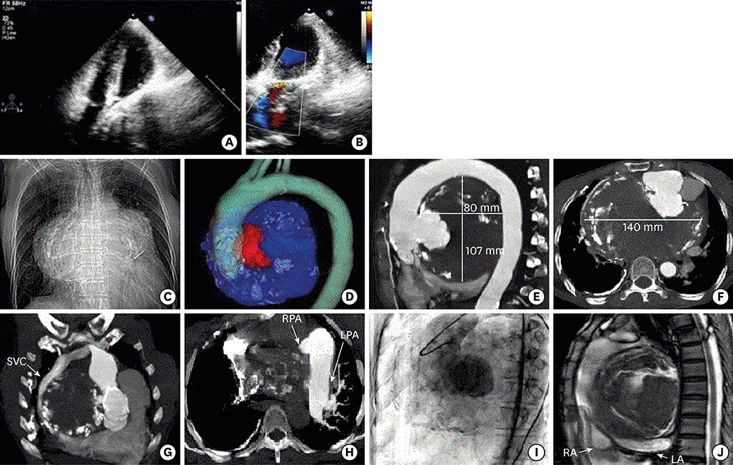
Figure 1 A & B - Transthoracic echocardiography – Extra cardiac mass compressing left atrium. C - Chest scanogram - lobular mediastinal widening with silhouetting of the ascending aorta. D - Volume rendered CT angiogram - Small patent lumen (600 HU) color-coded red and large peripheral thrombus (30 to 60 HU) color-coded blue. E & F - CT angiography - giant partially thrombosed aneurysm (80 × 107× 140 mm) arising from the posterior wall of the ascending aorta sinotubular junction. G - CT angiography - severely compressed patent SVC draping along right lateral aspect of aneurysm. H - Pulmonary arterial phase of CT Angiography - severely compressed Right Pulmonary artery. Mildly dilated opacified left Pulmonary artery. I - Aortography - saccular aneurysm of ascending aorta. J - Cardiac MRI (FIESTA) sagittal - variegated hypointense thrombus with severe inferior compression of the atria. LA: left atrium, LPA: left pulmonary artery, RA: right atrium, RPA: right pulmonary artery, SVC: superior vena cava.
- Full Text Links
-
- Actions
-
Cited
- CITED
-
- Close
- Share
-
- Similar articles
-
- A Case of Ascending Aortic Aneurysm Associated with Congenital Bicuspid Aortic Valve
- Aortic Valve Sparing Operations: A Review
- A Rare Congenital Anomaly: Interrupted Aortic Arch and Giant Ascending Aortic Aneurysm in a Young Male
- Sudden Cardiac Death from Acute Myocardial Infarction Caused by Unruptured Ascending Aortic Aneurysm Involving the Sinus of Valsalva: An Autopsy Case
- Abdominal aortic aneurysm in giant cell arteritis
