Pediatric Case with Acute Bilateral Serous Macular Detachment
- Affiliations
-
- 1Goznuru Eye Hospital, Gaziantep, Turkey.
- 2Dr. Ersin Arslan Education and Research Hospital, Gaziantep, Turkey. mustafaberhuni@gmail.com
- KMID: 2442625
- DOI: http://doi.org/10.3341/kjo.2018.0067
Abstract
- No abstract available.
MeSH Terms
Figure
-
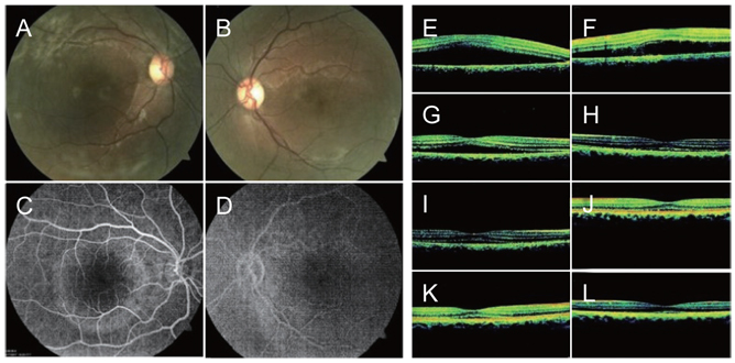
Fig. 1 (A,B) Fundus pictures showing the serous elevation in the right and left macula at presentation. (C,D) Leaking macular areas at the late phase in both eyes on fundus fluorescein angiography. (E,F) Optical coherence tomography (OCT) image showing serous macular detachment (SMD) in both eyes at presentation (right central macular thickness [CMT], 729 µm; left CMT, 663 µm). (G,H) OCT image showing decreased SMD at the fourth day follow-up (right CMT, 387 µm; left CMT, 273 µm). (I,J) OCT image showing significantly decreased SMD in both eyes at the thirteenth day follow-up (right CMT, 286 µm; left CMT, 260 µm). (K,L) OCT image showing almost complete resolution of the SMD in both eyes at the thirtieth day follow-up (right CMT, 267 µm; left CMT, 243 µm).
Reference
-
1. Regenbogen L, Stein R, Lazar M. Macular and juxtapapillar serous retinal detachment associated with pit of optic disc. Ophthalmologica. 1964; 148:247–251.
Article2. Tregoning JS, Schwarze J. Respiratory viral infections in infants: causes, clinical symptoms, virology, and immunology. Clin Microbiol Rev. 2010; 23:74–98.
Article3. Ovet G, Alpfidan I, Sakarya Y, et al. The acute effect of pseudoephedrine on choroidal thickness. Clin Ter. 2016; 167:63–66.4. Michael JC, Pak J, Pulido J, de Venecia G. Central serous chorioretinopathy associated with administration of sympathomimetic agents. Am J Ophthalmol. 2003; 136:182–185.
Article
- Full Text Links
-
- Actions
-
Cited
- CITED
-
- Close
- Share
-
- Similar articles
-
- Bilateral Serous Retinal Detachment Associated With Alport's Syndrome
- Laser Photocoaculation Treatment in a Case of Circumscribged Choroidal hmangioma Associated with Serous Retinal Detachment
- A Case of Optic Disc Pit
- Spontaneous Resolution of Post-Traumatic Bilateral Serous Retinal Detachment in Childrens
- A Case of Surgically Treated Serous Macular Detachment Associated With Optic Disc Pit
