Breast-Specific Gamma Imaging in Breast Cancer Screening
- Affiliations
-
- 1Department of Radiology, Ewha Womans University, School of Medicine, Seoul, Korea. escha@ewha.ac.kr
- KMID: 2442464
- DOI: http://doi.org/10.3348/jksr.2019.80.1.59
Abstract
- The sensitivity and specificity of breast cancer screening with mammography in dense breasts are very low, and increasing attention has been paid to breast density and screening in high-risk groups. Therefore, a supplemental screening modality is needed. Mammography and supplementary ultrasound screening are mainly based on the differences between cancerous and normal breast parenchyma on an anatomical basis, whereas breast-specific gamma imaging is a functional imaging approach examining the physiological phenomena of blood flow and mitochondrial activity, which are increased in cancer cells. The purpose of this review is to discuss the diagnostic methods, clinical results, clinical application, and considerations of breast cancer screening with breast-specific gamma imaging using (99m)Tc-sestamibi.
MeSH Terms
Figure
-
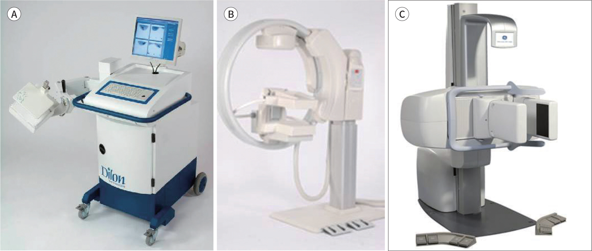
Fig. 1. Three different breast-specific gamma imaging systems. A. Dilon Diagnostics 6800 (Dilon Diagnostics). B. Gamma Medica LumaGEM 3200S (Gamma Medica Inc.). C. GE Healthcare Discovery NM750b (GE Healthcare).
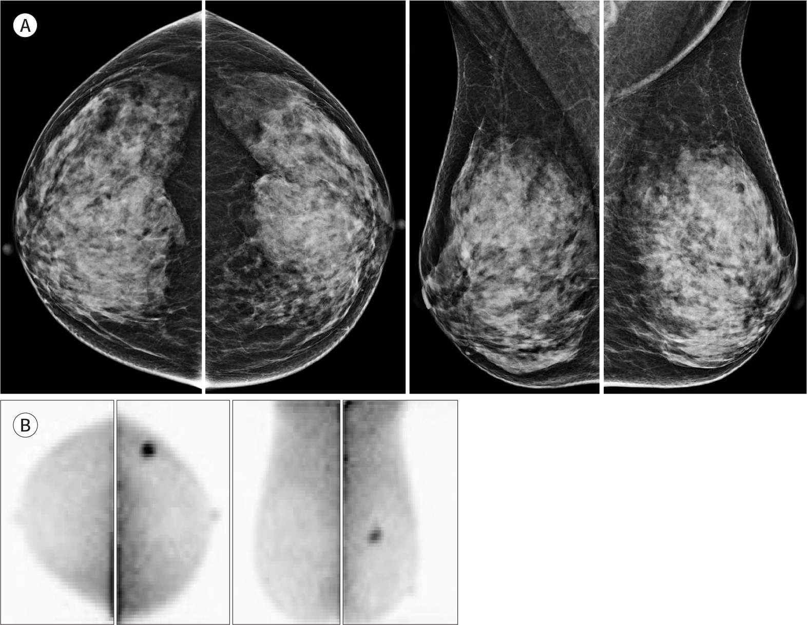
Fig. 2. A 48-year-old woman with invasive ductal carcinoma in left breast. A. Mammography of both breasts shows extremely dense breast without mass or microcalcification. B. Breast-specific gamma imaging shows abnormal focal intense uptake in the mid-outer region of the left breast.
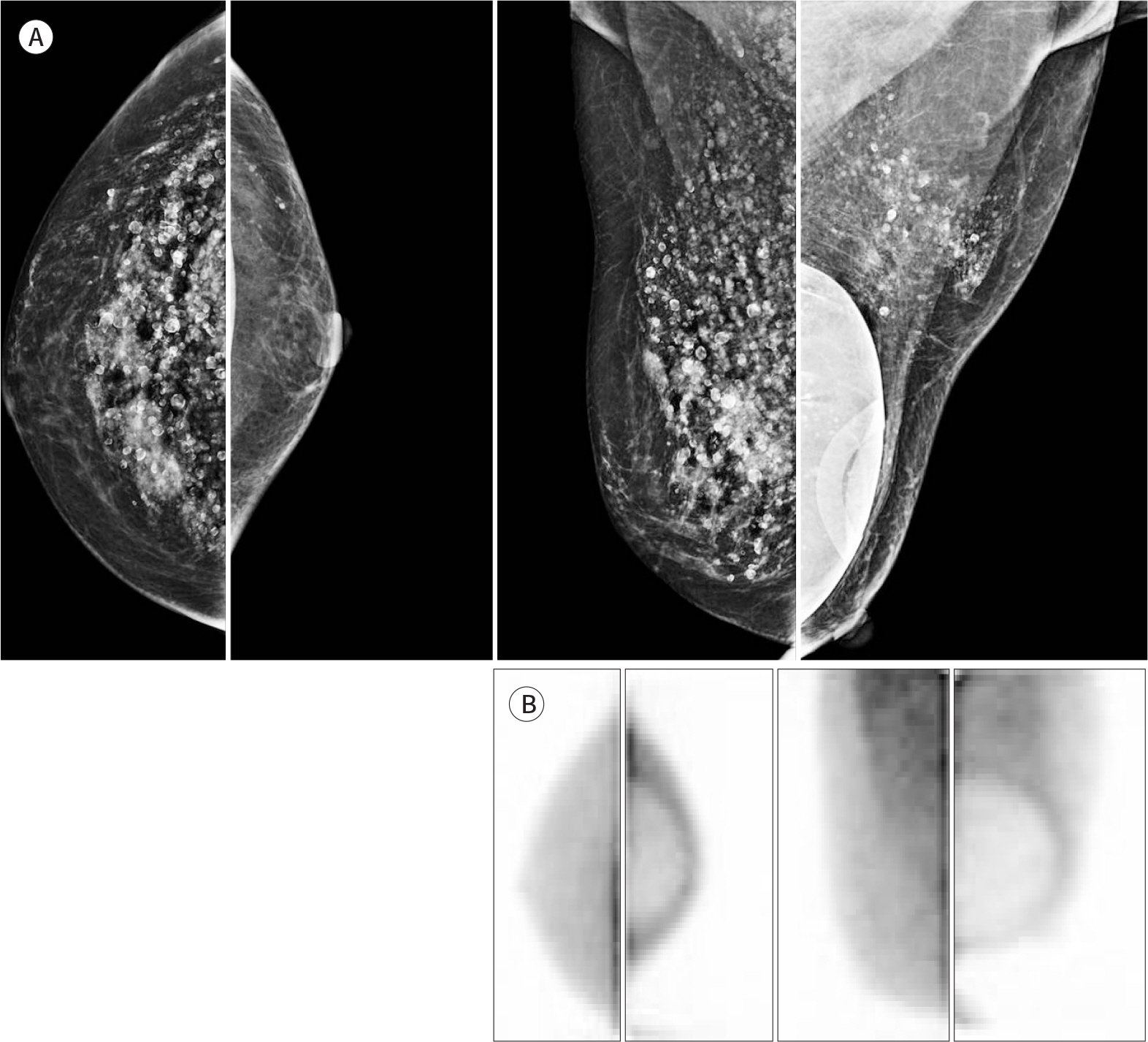
Fig. 3. A 68-year-old woman, underwent breast conserving surgery, with silicone injection mammoplasty and augmentation mammoplasty. A. Mammography of both breasts shows postsurgical deformity in the left breast with bilateral silicone granuloma and left mammoplasty bag. B. Breast-specific gamma imaging shows no abnormal uptake in both breasts.
Reference
-
References
1. Jung KW, Won YJ, Kong HJ, Oh CM, Cho H, Lee DH, et al. Cancer statistics in Korea: incidence, mortality, survival, and prevalence in 2012. Cancer Res Treat. 2015; 47:127–141.
Article2. Kim SY, Kim EK. Benefits and harms of breast screening: focus on updated Korean guideline for breast cancer screening. J Korean Soc Radiol. 2016; 74:147–155.3. Myers ER, Moorman P, Gierisch JM, Havrilesky LJ, Grimm LJ, Ghate S, et al. Benefits and harms of breast cancer screening: a systematic review.JAMA. 2015; 314:1615–1634.4. Sardanelli F, Podo F, D'Agnolo G, Verdecchia A, Santaquilani M, Musumeci R, et al. Multicenter comparative multimodality surveillance of women at genetic-familial high risk for breast cancer (HIBCRIT study): interim results. Radiology. 2007; 242:698–715.
Article5. Boyd NF, Guo H, Martin LJ, Sun L, Stone J, Fishell E, et al. Mammographic density and the risk and detection of breast cancer. N Engl J Med. 2007; 356:227–236.
Article6. Kerlikowske K, Cook AJ, Buist DS, Cummings SR, Vachon C, Vacek P, et al. Breast cancer risk by breast density, menopause, and postmenopausal hormone therapy use.J Clin Oncol. 2010; 28:3830–3837.7. Ubel PA. Medical facts versus value judgments–toward preference-sensitive guidelines.N Engl J Med. 2015; 372:2475–2477.8. Moon YK. Primary modality of breast ultrasonography in breast screening.J Br Screening. 2016; 13:83–87.9. Sharma S, Sharma MC, Sarkar C. Morphology of angiogenesis in human cancer: a conceptual overview, histoprognostic perspective and significance of neoangiogenesis.Histopathology. 2005; 46:481–489.10. Delmon-Moingeon LI, Piwnica-Worms D, Van den Abbeele AD, Holman BL, Davison A, Jones AG. Uptake of the cation hexakis(2-methyoxyisobutylisonitrile)-technetium-99m by human carcinoma cell lines in vitro. Cancer Res. 1990; 50:2198–2202.11. Sampalis FS, Denis R, Picard D, Fleiszer D, Martin G, Nassif E, et al. International prospective evaluation of scintimammography with (99m)technetium sestamibi.Am J Surg. 2003; 185:544–549.12. Zhou M, Johnson N, Gruner S, Ecklund GW, Meunier P, Bryn S, et al. Clinical utility of breast-specific gamma imaging for evaluating disease extent in the newly diagnosed breast cancer patient.Am J Surg. 2009; 197:159–163.13. Berg WA. Nuclear breast imaging: clinical results and future directions.J Nucl Med. 2016; 57(Suppl 1):46S–52S.14. Hsu DF, Freese DL, Levin CS. Breast-dedicated radionuclide imaging systems.J Nucl Med. 2016; 57(Suppl 1):40S–45S.15. Huppe AI, Mehta AK, Brem RF. Molecular breast imaging: a comprehensive review.Semin Ultrasound CT MR. 2018; 39:60–69.16. Khalkhali I, Mena I, Jouanne E, Diggles L, Venegas R, Block J, et al. Prone scintimammography in patients with suspicion of carcinoma of the breast.J Am Coll Surg. 1994; 178:491–497.17. Shermis RB, Redfern RE, Burns J, Kudrolli H. Molecular breast imaging in breast cancer screening and problem solving.Radiographics. 2017; 37:1309–1327.18. Narayanan D, Berg WA. Dedicated breast gamma camera imaging and breast PET: current status and future directions.PET Clin. 2018; 13:363–381.19. Khalkhali I, Iraniha S, Diggles LE, Cutrone JA, Mishkin FS. Scintimammography: the new role of technetium-99m Sestamibi imaging for the diagnosis of breast carcinoma. Q J Nucl Med. 1997; 41:231–238.20. Avril N, Rosé CA, Schelling M, Dose J, Kuhn W, Bense S, et al. Breast imaging with positron emission tomography and fluorine-18 fluorodeoxyglucose: use and limitations. J Cl/iin Oncol. 2000; 18:3495–3502.
Article21. O'Connor MK, Hruska CB, Tran TD, Swanson T, Conners AL, Jones K, et al. Factors influencing the uptake of 99mTc-sestamibi in breast tissue on molecular breast imaging.J Med Nucl Technol. 2015; 43:13–20.22. Rhodes DJ, Hruska CB, Phillips SW, Whaley DH, O'Connor MK. Dedicated dual-head gamma imaging for breast cancer screening in women with mammographically dense breasts. Radiology. 2011; 258:106–118.
Article23. Rhodes DJ, Hruska CB, Conners AL, Tortorelli CL, Maxwell RW, Jones KN, et al. Molecular breast imaging at reduced radiation dose for supplemental screening in mammographically dense breasts.AJR Am J Roentgenol. 2015; 204:241–251.24. Hruska CB. Molecular breast imaging for screening in dense breasts: state of the art and future directions. AJR Am J Roentgenol. 2017; 208:275–283.
Article25. Lee A, Chang J, Lim W, Kim BS, Lee JE, Cha ES, et al. Effectiveness of breast-specific gamma imaging (BSGI) for breast cancer in Korea: a comparative study. Breast J. 2012; 18:453–458.
Article26. Park JY, Yi SY, Park HJ, Kim MS, Kwon HJ, Park NH, et al. Breast-specific gamma imaging: correlations with mammographic and clinicopathologic characteristics of breast cancer. AJR Am J Roentgenol. 2014; 203:223–228.
Article27. Shermis RB, Wilson KD, Doyle MT, Martin TS, Merryman D, Kudrolli H, et al. Supplemental breast cancer screening with molecular breast imaging for women with dense breast tissue.AJR Am J Roentgenol. 2016; 207:450–457.28. Rechtman LR, Lenihan MJ, Lieberman JH, Teal CB, Torrente J, Rapelyea JA, et al. Breast-specific gamma imaging for the detection of breast cancer in dense versus nondense breasts.AJR Am J Roentgenol. 2014; 202:293–298.29. Hruska CB, Conners AL, Jones KN, O'Connor MK, Moriarty JP, Boughey JC, et al. Diagnostic workup and costs of a single supplemental molecular breast imaging screen of mammographically dense breasts. AJR Am J Roentgenol. 2015; 204:1345–1353.
Article30. Weigert JM, Bertrand ML, Lanzkowsky L, Stern LH, Kieper DA. Results of a multicenter patient registry to determine the clinical impact of breast-specific gamma imaging, a molecular breast imaging technique. AJR Am J Roentgenol. 2012; 198:W69–W75.
Article31. Kuhn KJ, Rapelyea JA, Torrente J, Teal CB, Brem RF. Comparative diagnostic utility of low-dose breast specific gamma imaging to current clinical standard.Breast J. 2016; 22:180–188.32. Kim BS. Usefulness of breast-specific gamma imaging as an adjunct modality in breast cancer patients with dense breast: a comparative study with MRI. Ann Nucl Med. 2012; 26:131–137.
Article33. Hendrick RE. Radiation doses and cancer risks from breast imaging studies. Radiology. 2010; 257:246–253.
Article34. Hendrick RE, Pisano ED, Averbukh A, Moran C, Berns EA, Yaffe MJ, et al. Comparison of acquisition parameters and breast dose in digital mammography and screen-film mammography in the American College of Radiology Imaging Network digital mammographic imaging screening trial.AJR Am J Roentgenol. 2010; 194:362–369.35. O'Connor MK, Li H, Rhodes DJ, Hruska CB, Clancy CB, Vetter RJ. Comparison of radiation exposure and associated radiation-induced cancer risks from mammography and molecular imaging of the breast.Med Phys. 2010; 37:6187–6198.36. Hendrick RE, Tredennick T. Benefit to radiation risk of breast specific gamma imaging compared with mammography in screening asymptomatic women with dense breasts.Radiology. 2016; 281:583–588.37. Brem RF, Fishman M, Rapelyea J. Young H, Teal C, Kelly T. Breast specific gamma imaging with Tc-sestamibi and magnetic resonance imaging in the diagnosis of breast cancer –a comparative study.Breast J. 2007; 13:465–469.38. National Comprehensive Cancer Network. NCCN guidelines for detection, prenvention,& risk reduction. Available at:. https://www.nccn.org/professionals/physician_gls/. Accessed Dec 1,. 2018.39. Goldsmith SJ, Parsons W, Guiberteau MJ, Stern LH, Lanzkowsky L, Weigert J, et al. SNM practice guideline for breast scintigraphy with breast-specific gamma-cameras 1.0. J Nucl Med Technol. 2010; 38:219–224.40. Lee SJ, Choi YY, Kim C, Chung MS. Correlations between tumor to background ratio on breast-specific gamma imaging and prognostic factors in breast cancer. J Korean Med Sci. 2017; 32:1031–1037.
Article
