Prediction of Pulmonary Function in Patients with Chronic Obstructive Pulmonary Disease: Correlation with Quantitative CT Parameters
- Affiliations
-
- 1Department of Radiology and Research Institute of Radiology, Asan Medical Center, University of Ulsan College of Medicine, Seoul, Korea. asellion@hanmail.net
- 2Department of Convergence Medicine, Asan Medical Center, University of Ulsan College of Medicine, Seoul, Korea.
- 3Department of Pulmonary and Critical Care Medicine and Clinical Research Center for Chronic Obstructive Airway Diseases, Asan Medical Center, University of Ulsan College of Medicine, Seoul, Korea.
- KMID: 2440491
- DOI: http://doi.org/10.3348/kjr.2018.0391
Abstract
OBJECTIVE
We aimed to evaluate correlations between computed tomography (CT) parameters and pulmonary function test (PFT) parameters according to disease severity in patients with chronic obstructive pulmonary disease (COPD), and to determine whether CT parameters can be used to predict PFT indices.
MATERIALS AND METHODS
A total of 370 patients with COPD were grouped based on disease severity according to the Global Initiative for Chronic Obstructive Lung Disease (GOLD) I-IV criteria. Emphysema index (EI), air-trapping index, and airway parameters such as the square root of wall area of a hypothetical airway with an internal perimeter of 10 mm (Pi10) were measured using automatic segmentation software. Clinical characteristics including PFT results and quantitative CT parameters according to GOLD criteria were compared using ANOVA. The correlations between CT parameters and PFT indices, including the ratio of forced expiratory volume in one second to forced vital capacity (FEV1/FVC) and FEV1, were assessed. To evaluate whether CT parameters can be used to predict PFT indices, multiple linear regression analyses were performed for all patients, Group 1 (GOLD I and II), and Group 2 (GOLD III and IV).
RESULTS
Pulmonary function deteriorated with increase in disease severity according to the GOLD criteria (p < 0.001). Parenchymal attenuation parameters were significantly worse in patients with higher GOLD stages (p < 0.001), and Pi10 was highest for patients with GOLD III (4.41 ± 0.94 mm). Airway parameters were nonlinearly correlated with PFT results, and Pi10 demonstrated mild correlation with FEV1/FVC in patients with GOLD II and III (r = 0.16, p = 0.06 and r = 0.21, p = 0.04, respectively). Parenchymal attenuation parameters, airway parameters, EI, and Pi10 were identified as predictors of FEV1/FVC for the entire study sample and for Group 1 (R2 = 0.38 and 0.22, respectively; p < 0.001). However, only parenchymal attenuation parameter, EI, was identified as a predictor of FEV1/FVC for Group 2 (R2 = 0.37, p < 0.001). Similar results were obtained for FEV1.
CONCLUSION
Airway and parenchymal attenuation parameters are independent predictors of pulmonary function in patients with mild COPD, whereas parenchymal attenuation parameters are dominant independent predictors of pulmonary function in patients with severe COPD.
Keyword
MeSH Terms
Figure
-
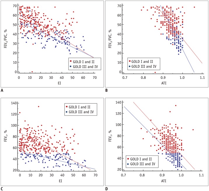
Fig. 1 Scatter plots of correlations between EI (A, C) or ATI (B, D) and pulmonary function indices.Scatter plots of distributions of four GOLD stages are included in Supplementary Figure 1 (in the online-only Data Supplement). ATI = air-trapping index, EI = emphysema index, FEV1 = percentage predicted forced expiratory volume in one second, FEV1/FVC = ratio of forced expiratory volume in one second to forced vital capacity, GOLD = Global Initiative for Chronic Obstructive Lung Disease
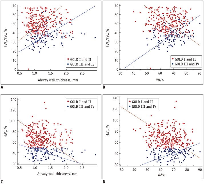
Fig. 2 Scatter plots of correlations of airway wall thickness (A, C) and WA% (B, D) with pulmonary function indices.Scatter plots of distributions of four GOLD stages are included in Supplementary Figure 2 (in the online-only Data Supplement). WA% = wall area percentage
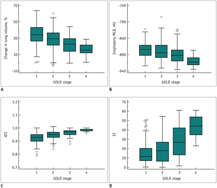
Fig. 3 Distribution of CT parameters according to GOLD stages.(A) Changes in lung volume ([inspiratory volume − expiratory volume] / inspiratory volume × 100) in whole lungs, (B) inspiratory MLD, (C) ATI (ratio of MLD on expiration and inspiration), and (D) EI measured on inspiration CT according to GOLD stages. Change in lung volume is smaller for more advanced GOLD stages. Inspiratory MLD as well as ATI decrease, whereas EI increases, with increase in GOLD stage. CT = computed tomography, HU = Hounsfield unit, MLD = mean lung density
Reference
-
1. Vogelmeier CF, Criner GJ, Martinez FJ, Anzueto A, Barnes PJ, Bourbeau J, et al. Global strategy for the diagnosis, management, and prevention of chronic obstructive lung disease 2017 report. GOLD executive summary. Am J Respir Crit Care Med. 2017; 195:557–582. PMID: 28128970.
Article2. Topalovic M, Laval S, Aerts JM, Troosters T, Decramer M, Janssens W. Automated interpretation of pulmonary function tests in adults with respiratory complaints. Respiration. 2017; 93:170–178. PMID: 28088797.
Article3. Al-Ashkar F, Mehra R, Mazzone PJ. Interpreting pulmonary function tests: recognize the pattern, and the diagnosis will follow. Cleve Clin J Med. 2003; 70:866868871–873. passim. PMID: 14621232.
Article4. Lynch DA, Al-Qaisi MA. Quantitative computed tomography in chronic obstructive pulmonary disease. J Thorac Imaging. 2013; 28:284–290. PMID: 23748651.
Article5. Schroeder JD, McKenzie AS, Zach JA, Wilson CG, Curran-Everett D, Stinson DS, et al. Relationships between airflow obstruction and quantitative CT measurements of emphysema, air trapping, and airways in subjects with and without chronic obstructive pulmonary disease. AJR Am J Roentgenol. 2013; 201:W460–W470. PMID: 23971478.
Article6. Paoletti M, Cestelli L, Bigazzi F, Camiciottoli G, Pistolesi M. Chronic obstructive pulmonary disease: pulmonary function and CT lung attenuation do not show linear correlation. Radiology. 2015; 276:571–578. PMID: 25848902.
Article7. Park TS, Lee JS, Seo JB, Hong Y, Yoo JW, Kang BJ, et al. Study design and outcomes of Korean Obstructive Lung Disease (KOLD) cohort study. Tuberc Respir Dis (Seoul). 2014; 76:169–174. PMID: 24851130.
Article8. Ekberg-Aronsson M, Pehrsson K, Nilsson JA, Nilsson PM, Löfdahl CG. Mortality in GOLD stages of COPD and its dependence on symptoms of chronic bronchitis. Respir Res. 2005; 6:98. PMID: 16120227.
Article9. Quanjer PH, Tammeling GJ, Cotes JE, Pedersen OF, Peslin R, Yernault JC. Lung volumes and forced ventilatory flows. Report working party standardization of lung function tests, European community for steel and coal. Official statement of the European Respiratory Society. Eur Respir J Suppl. 1993; 16:5–40. PMID: 8499054.10. American Thoracic Society. Single-breath carbon monoxide diffusing capacity (transfer factor). Recommendations for a standard technique--1995 update. Am J Respir Crit Care Med. 1995; 152:2185–2198. PMID: 8520796.11. Standardization of spirometry, 1994 Update. Am J Respir Crit Care Med. 1995; 152:1107–1136. PMID: 7663792.12. Cho YH, Seo JB, Kim N, Lee HJ, Hwang HJ, Kim EY, et al. Comparison of a new integral-based half-band method for CT measurement of peripheral airways in COPD with a conventional full-width half-maximum method using both phantom and clinical CT images. J Comput Assist Tomogr. 2015; 39:428–436. PMID: 25700223.
Article13. Cerveri I, Dore R, Corsico A, Zoia MC, Pellegrino R, Brusasco V, et al. Assessment of emphysema in COPD: a functional and radiologic study. Chest. 2004; 125:1714–1718. PMID: 15136381.14. Agusti A, Calverley PM, Celli B, Coxson HO, Edwards LD, Lomas DA, et al. Characterisation of COPD heterogeneity in the ECLIPSE cohort. Respir Res. 2010; 11:122. PMID: 20831787.
Article15. Bafadhel M, Umar I, Gupta S, Raj JV, Vara DD, Entwisle JJ, et al. The role of CT scanning in multidimensional phenotyping of COPD. Chest. 2011; 140:634–642. PMID: 21454400.
Article16. Lynch DA, Austin JH, Hogg JC, Grenier PA, Kauczor HU, Bankier AA, et al. CT-definable subtypes of chronic obstructive pulmonary disease: a statement of the Fleischner society. Radiology. 2015; 277:192–205. PMID: 25961632.
Article17. Smith BM, Austin JH, Newell JD Jr, D'Souza BM, Rozenshtein A, Hoffman EA, et al. Pulmonary emphysema subtypes on computed tomography: the MESA COPD study. Am J Med. 2014; 127:94.e7–94.e23.
Article18. Bhatt SP, Sieren JC, Dransfield MT, Washko GR, Newell JD Jr, Stinson DS, et al. Comparison of spirometric thresholds in diagnosing smoking-related airflow obstruction. Thorax. 2014; 69:409–414. PMID: 23525095.
Article19. Donohue KM, Hoffman EA, Baumhauer H, Guo J, Budoff M, Austin JH, et al. Cigarette smoking and airway wall thickness on CT scan in a multi-ethnic cohort: the MESA Lung Study. Respir Med. 2012; 106:1655–1664. PMID: 22974831.
Article20. Kohansal R, Soriano JB, Agusti A. Investigating the natural history of lung function: facts, pitfalls, and opportunities. Chest. 2009; 135:1330–1341. PMID: 19420200.21. Oga T, Tsukino M, Hajiro T, Ikeda A, Koyama H, Mishima M, et al. Multidimensional analyses of long-term clinical courses of asthma and chronic obstructive pulmonary disease. Allergol Int. 2010; 59:257–265. PMID: 20657164.
Article22. Galbán CJ, Han MK, Boes JL, Chughtai KA, Meyer CR, Johnson TD, et al. Computed tomography-based biomarker provides unique signature for diagnosis of COPD phenotypes and disease progression. Nat Med. 2012; 18:1711–1715. PMID: 23042237.
Article23. McDonough JE, Yuan R, Suzuki M, Seyednejad N, Elliott WM, Sanchez PG, et al. Small-airway obstruction and emphysema in chronic obstructive pulmonary disease. N Engl J Med. 2011; 365:1567–1575. PMID: 22029978.
Article24. Kim C, Lee KY, Shin C, Kang EY, Oh YW, Ha M, et al. Comparison of filtered back projection, hybrid iterative reconstruction, model-based iterative reconstruction, and virtual monoenergetic reconstruction images at both low- and standard-dose settings in measurement of emphysema volume and airway wall thickness: a CT phantom study. Korean J Radiol. 2018; 19:809–817. PMID: 29962888.
Article
- Full Text Links
-
- Actions
-
Cited
- CITED
-
- Close
- Share
-
- Similar articles
-
- Clinical use of chest CT in chronic obstructive pulmonary diseases
- Comparison of Inhalation Scan and Perfusion Scan for the Prediction of Postoperative Pulmonary Function
- Quantification of Emphysema with a Three-Dimensional Chest CT Scan: Correlation with the Visual Emphysema Scoring on Chest CT, Pulmonary Function Tests and Dyspnea Severity
- Quantitative CT Assessment in Chronic Obstructive Pulmonary Disease Patients: Comparison of the Patients with and without Consistent Clinical Symptoms and Pulmonary Function Results
- Quantitative CT Imaging in Chronic Obstructive Pulmonary Disease: Review of Current Status and Future Challenges
