Measurement of Pancreatic Fat Fraction by CT Histogram Analysis to Predict Pancreatic Fistula after Pancreaticoduodenectomy
- Affiliations
-
- 1Department of Radiology, Hallym University Sacred Heart Hospital, Anyang, Korea. ha.hongil@gmail.com
- 2Department of Surgery, Hallym University Sacred Heart Hospital, Anyang, Korea.
- KMID: 2440483
- DOI: http://doi.org/10.3348/kjr.2018.0557
Abstract
OBJECTIVE
To evaluate the effectiveness of computed tomography (CT) Hounsfield unit histogram analysis (HUHA) in postoperative pancreatic fistula (PF) prediction.
MATERIALS AND METHODS
Fifty-four patients (33 males and 21 females; mean age, 65.6 years; age range, 37-89 years) who had undergone preoperative CT and pancreaticoduodenectomy were included in this retrospective study. Two radiologists measured mean CT Hounsfield unit (CTHU) values by drawing regions of interest (ROIs) at the level of the pancreaticojejunostomy site on preoperative pre-contrast images. The HUHA values were arbitrarily divided into three categories, comprising HUHA-A ≤ 0 HU, 0 HU < HUHA-B < 30 HU, and HUHA-C ≥ 30 HU. Each HUHA value within the ROI was calculated as a percentage of the entire area using commercial 3-dimensional analysis software. Pancreas texture was evaluated as soft or hard by manual palpation.
RESULTS
Fifteen patients (27.8%) had clinically relevant PFs. The PF group had significantly higher HUHA-A (p < 0.01) and significantly lower mean CTHU (p < 0.01) values than those of the non-PF group. The HUHA-A value had a moderately strong correlation with PF occurrence (r = 0.60, p < 0.01), whereas the mean CTHU had a weak negative correlation with PF occurrence (r = −0.27, p < 0.01). The HUHA-A and mean CTHU areas under the curve (AUCs) for predicting PF occurrence were 0.86 and 0.65, respectively, with significant difference (p < 0.01). The HUHA-A and mean CTHU AUCs for predicting pancreatic softness were 0.86 and 0.64, respectively, with significant difference (p < 0.01).
CONCLUSION
The HUHA-A values on preoperative pre-contrast CT images demonstrate a strong correlation with PF occurrence.
Keyword
MeSH Terms
Figure
-
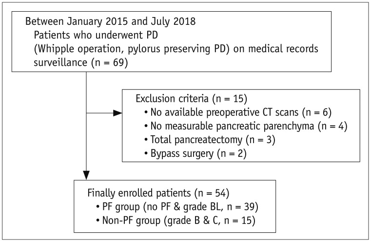
Fig. 1 Patient selection flowchart.BL = biochemical fistula, CT = computed tomography, PD = pancreaticoduodenectomy, PF = pancreatic fistula
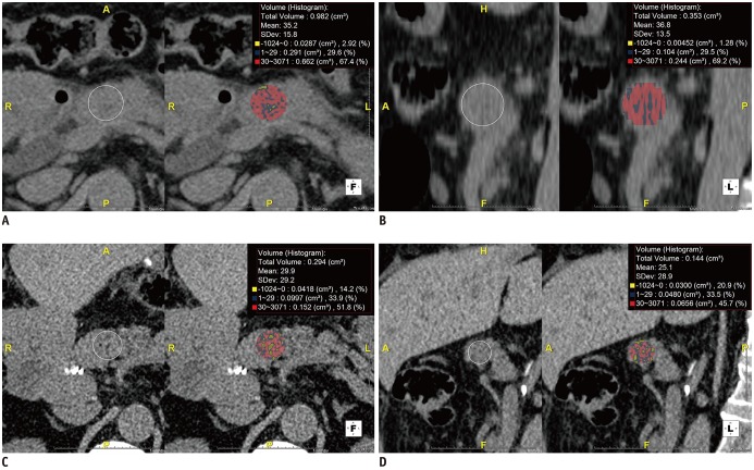
Fig. 2 Measurement of HUHA.ROIs were drawn on axial and corresponding sagittal preoperative pre-contrast CT images at level of pancreas neck near pancreaticojejunostomy site with reference to postoperative CT images. Three-dimensional analysis software automatically generated each HUHA-A (−1024–0 HU), HUHA-B (1–29 HU), HUHA-C (30–3071 HU), and mean CTHU value. Each HUHA value within ROI was calculated as percentage of entire area in real time.Axial (A) and sagittal (B) images from 76-year-old male patient without PF who underwent pylorus-preserving PD for pancreatic cancer show HUHA-A of 2.1% and mean CTHU of 36 HU. Axial (C) and sagittal (D) images from 77-year-old female patient with grade B PF who underwent pylorus-preserving PD for ampulla of Vater cancer show HUHA-A of 17.6% and mean CTHU of 27.5 HU. CTHU = CT Hounsfield unit, HUHA = Hounsfield unit histogram analysis, ROI = region of interest, SD = standard deviation
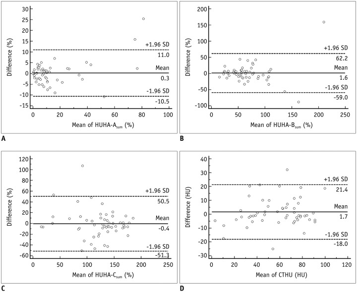
Fig. 3 Bland-Altman plots of inter-observer agreement for HUHA and mean CTHU measurements.A-D. Differences (y axis) between two observers are plotted against mean value (x axis) of their measurements. 95% limits of agreements of HUHA-A (A), HUHA-B (B), HUHA-C (C), and mean CTHU (D) ranged from −10.5% to 11% (mean, 0.3%), from −59% to 62.2% (mean, 1.6%), from −51.3% to 50.5% (mean −0.4%), and from −18.0 HU to 21.4 HU (mean 1.7 HU), respectively. Solid line indicates mean difference. Top and bottom dashed lines correspond to upper and lower margins of 95% limits of agreement. With probability of 95%, differences in normalized scores of future examinations will be between upper and lower limits of agreement (mean ± variability estimate = 1.96 × SD).
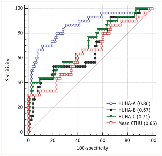
Fig. 4 ROC curve to predict PF using HUHA-A (AUC = 0.86), HUHA-B (AUC = 0.67), HUHA-C (AUC = 0.71), and mean CTHU (AUC = 0.65) values.AUC = area under curve, ROC = receiver operating characteristic
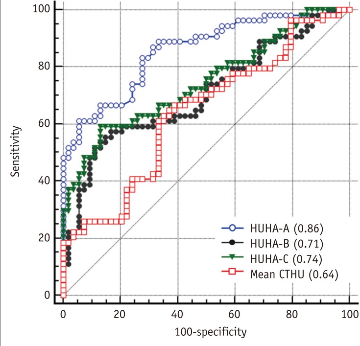
Fig. 5 ROC curve to predict pancreatic softness using HUHA-A (AUC = 0.86), HUHA-B (AUC = 0.71), HUHA-C (AUC = 0.74), and mean CTHU (AUC = 0.64) values.
Cited by 1 articles
-
The effect of atrophied pancreas as shown in the preoperative imaging on the leakage rate after pancreaticoduodenectomy
Ashraf Imam, Harbi Khalayleh, Meni Brakha, Ariel A. Benson, Naama Lev-Cohain, Gidon Zamir, Abed Khalaileh
Ann Hepatobiliary Pancreat Surg. 2022;26(2):184-189. doi: 10.14701/ahbps.21-145.
Reference
-
1. Lerut JP, Gianello PR, Otte JB, Kestens PJ. Pancreaticoduodenal resection. Surgical experience and evaluation of risk factors in 103 patients. Ann Surg. 1984; 199:432–437. PMID: 6712319.2. Yeo CJ, Cameron JL, Sohn TA, Lillemoe KD, Pitt HA, Talamini MA, et al. Six hundred fifty consecutive pancreaticoduodenectomies in the 1990s: pathology, complications, and outcomes. Ann Surg. 1997; 226:248–260. discussion 257-260. PMID: 9339931.3. Seo JW, Hwang HK, Lee M, Kim KW, Kang CM, Kim MJ, et al. Normal postoperative computed tomography findings after a variety of pancreatic surgeries. Korean J Radiol. 2017; 18:299–308. PMID: 28246510.
Article4. Miedema BW, Sarr MG, van Heerden JA, Nagorney DM, McIlrath DC, Ilstrup D. Complications following pancreaticoduodenectomy. Current management. Arch Surg. 1992; 127:945–950. discussion 949-950. PMID: 1353671.5. Vanounou T, Pratt WB, Callery MP, Vollmer CM Jr. Selective administration of prophylactic octreotide during pancreaticoduodenectomy: a clinical and cost-benefit analysis in low- and high-risk glands. J Am Coll Surg. 2007; 205:546–557. PMID: 17903728.
Article6. Roberts KJ, Storey R, Hodson J, Smith AM, Morris-Stiff G. Pre-operative prediction of pancreatic fistula: is it possible? Pancreatology. 2013; 13:423–428. PMID: 23890142.
Article7. van Berge Henegouwen MI, De Wit LT, Van Gulik TM, Obertop H, Gouma DJ. Incidence, risk factors, and treatment of pancreatic leakage after pancreaticoduodenectomy: drainage versus resection of the pancreatic remnant. J Am Coll Surg. 1997; 185:18–24. PMID: 9208956.
Article8. Bassi C, Dervenis C, Butturini G, Fingerhut A, Yeo C, Izbicki J, et al. Postoperative pancreatic fistula: an international study group (ISGPF) definition. Surgery. 2005; 138:8–13. PMID: 16003309.
Article9. Mathur A, Pitt HA, Marine M, Saxena R, Schmidt CM, Howard TJ, et al. Fatty pancreas: a factor in postoperative pancreatic fistula. Ann Surg. 2007; 246:1058–1064. PMID: 18043111.10. Bassi C, Marchegiani G, Dervenis C, Sarr M, Abu Hilal M, Adham M, et al. The 2016 update of the international study group (ISGPS) definition and grading of postoperative pancreatic fistula: 11 years after. Surgery. 2017; 161:584–591. PMID: 28040257.11. Rosso E, Casnedi S, Pessaux P, Oussoultzoglou E, Panaro F, Mahfud M, et al. The role of “fatty pancreas” and of BMI in the occurrence of pancreatic fistula after pancreaticoduodenectomy. J Gastrointest Surg. 2009; 13:1845–1851. PMID: 19639369.
Article12. Lee SE, Jang JY, Lim CS, Kang MJ, Kim SH, Kim MA, et al. Measurement of pancreatic fat by magnetic resonance imaging: predicting the occurrence of pancreatic fistula after pancreatoduodenectomy. Ann Surg. 2010; 251:932–936. PMID: 20395858.13. Sugimoto M, Takahashi S, Kojima M, Kobayashi T, Gotohda N, Konishi M. In patients with a soft pancreas, a thick parenchyma, a small duct, and fatty infiltration are significant risks for pancreatic fistula after pancreaticoduodenectomy. J Gastrointest Surg. 2017; 21:846–854. PMID: 28101719.
Article14. Machado NO. Pancreatic fistula after pancreatectomy: definitions, risk factors, preventive measures, and management-review. Int J Surg Oncol. 2012; 2012:602478. PMID: 22611494.15. Yardimci S, Kara YB, Tuney D, Attaallah W, Ugurlu MU, Dulundu E, et al. A simple method to evaluate whether pancreas texture can be used to predict pancreatic fistula risk after pancreatoduodenectomy. J Gastrointest Surg. 2015; 19:1625–1631. PMID: 25982120.
Article16. Lim HK, Ha HI, Park SY, Lee K. Comparison of the diagnostic performance of CT Hounsfield unit histogram analysis and dual-energy X-ray absorptiometry in predicting osteoporosis of the femur. Eur Radiol. 2018; 9. 25. DOI: 10.1007/s00330-018-5728-0. [Epub ahead of print].
Article17. Hu BY, Wan T, Zhang WZ, Dong JH. Risk factors for postoperative pancreatic fistula: analysis of 539 successive cases of pancreaticoduodenectomy. World J Gastroenterol. 2016; 22:7797–7805. PMID: 27678363.
Article18. McMillan MT, Vollmer CM Jr, Asbun HJ, Ball CG, Bassi C, Beane JD, et al. The characterization and prediction of ISGPF grade C fistulas following pancreatoduodenectomy. J Gastrointest Surg. 2016; 20:262–276. PMID: 26162925.
Article19. Hashimoto Y, Sclabas GM, Takahashi N, Kirihara Y, Smyrk TC, Huebner M, et al. Dual-phase computed tomography for assessment of pancreatic fibrosis and anastomotic failure risk following pancreatoduodenectomy. J Gastrointest Surg. 2011; 15:2193–2204. PMID: 21948179.
Article20. Frozanpor F, Loizou L, Ansorge C, Lundell L, Albiin N, Segersvärd R. Correlation between preoperative imaging and intraoperative risk assessment in the prediction of postoperative pancreatic fistula following pancreatoduodenectomy. World J Surg. 2014; 38:2422–2429. PMID: 24711156.
Article21. DeLong ER, DeLong DM, Clarke-Pearson DL. Comparing the areas under two or more correlated receiver operating characteristic curves: a nonparametric approach. Biometrics. 1988; 44:837–845. PMID: 3203132.
Article22. Park SH, Goo JM, Jo CH. Receiver operating characteristic (ROC) curve: practical review for radiologists. Korean J Radiol. 2004; 5:11–18. PMID: 15064554.
Article23. Kim SY, Kim H, Cho JY, Lim S, Cha K, Lee KH, et al. Quantitative assessment of pancreatic fat by using unenhanced CT: pathologic correlation and clinical implications. Radiology. 2014; 271:104–112. PMID: 24475851.
Article24. Pickhardt PJ, Graffy PM, Reeder SB, Hernando D, Li K. Quantification of liver fat content with unenhanced MDCT: phantom and clinical correlation with MRI proton density fat fraction. AJR Am J Roentgenol. 2018; 211:W151–W157. PMID: 30016142.
Article25. Kramer H, Pickhardt PJ, Kliewer MA, Hernando D, Chen GH, Zagzebski JA, et al. Accuracy of liver fat quantification with advanced CT, MRI, and ultrasound techniques: prospective comparison with MR spectroscopy. AJR Am J Roentgenol. 2017; 208:92–100. PMID: 27726414.
Article26. Yoon JH, Lee JM, Lee KB, Kim SW, Kang MJ, Jang JY, et al. Pancreatic steatosis and fibrosis: quantitative assessment with preoperative multiparametric MR imaging. Radiology. 2016; 279:140–150. PMID: 26566228.
Article27. Kawai M, Kondo S, Yamaue H, Wada K, Sano K, Motoi F, et al. Predictive risk factors for clinically relevant pancreatic fistula analyzed in 1,239 patients with pancreaticoduodenectomy: multicenter data collection as a project study of pancreatic surgery by the Japanese Society of Hepato-Biliary-Pancreatic Surgery. J Hepatobiliary Pancreat Sci. 2011; 18:601–608. PMID: 21491103.
Article28. Sugimoto M, Takahashi S, Kojima M, Gotohda N, Kato Y, Kawano S, et al. What is the nature of pancreatic consistency? Assessment of the elastic modulus of the pancreas and comparison with tactile sensation, histology, and occurrence of postoperative pancreatic fistula after pancreaticoduodenectomy. Surgery. 2014; 156:1204–1211. PMID: 25444318.
Article29. Chang YR, Kang JS, Jang JY, Jung WH, Kang MJ, Lee KB, et al. Prediction of pancreatic fistula after distal pancreatectomy based on cross-sectional images. World J Surg. 2017; 41:1610–1617. PMID: 28091744.
Article30. Sugimoto M, Takahashi S, Gotohda N, Kato Y, Kinoshita T, Shibasaki H, et al. Schematic pancreatic configuration: a risk assessment for postoperative pancreatic fistula after pancreaticoduodenectomy. J Gastrointest Surg. 2013; 17:1744–1751. PMID: 23975030.
Article31. Okano K, Oshima M, Kakinoki K, Yamamoto N, Akamoto S, Yachida S, et al. Pancreatic thickness as a predictive factor for postoperative pancreatic fistula after distal pancreatectomy using an endopath stapler. Surg Today. 2013; 43:141–147. PMID: 22782593.
Article32. Pompe E, Willemink MJ, Dijkhuis GR, Verhaar HJ, Mohamed Hoesein FA, de Jong PA. Intravenous contrast injection significantly affects bone mineral density measured on CT. Eur Radiol. 2015; 25:283–289. PMID: 25187384.
Article33. Bushberg JT, Seibert JA, Leidholdt EM Jr, Boone JM. The essential physics of medical imaging. 3rd ed. Philadelphia, PA: Wolters Kluwer Health/Lippincott Williams and Wilkins;2012. p. 324.34. Tempero MA, Arnoletti JP, Behrman SW, Ben-Josef E, Benson AB 3rd, Casper ES, et al. Pancreatic Adenocarcinoma, version 2.2012: featured updates to the NCCN Guidelines. J Natl Compr Canc Netw. 2012; 10:703–713. PMID: 22679115.
- Full Text Links
-
- Actions
-
Cited
- CITED
-
- Close
- Share
-
- Similar articles
-
- A model for predicting pancreatic leakage after pancreaticoduodenectomy based on the international study group of pancreatic surgery classification
- The Efficacy of the Prophylactic Use of Octreotide after a Pancreaticoduodenectomy
- The impact of perioperative inotropes on the incidence of pancreatic leak following pancreaticoduodenectomy
- Factors Influencing the Pancreatic Leakage after Pancreaticoduodenectomy
- A Clinical Analysis on the Pancreaticoduodenectomy
